Clinical Trial
. 2022 Mar:6:e2100239.
doi: 10.1200/PO.21.00239.

Hyperthermic Intraperitoneal Chemotherapy-Induced Molecular Changes in Humans Validate Preclinical Data in Ovarian Cancer

Affiliations
Clinical Trial

Hyperthermic Intraperitoneal Chemotherapy-Induced Molecular Changes in Humans Validate Preclinical Data in Ovarian Cancer

Thanh H Dellinger et al. JCO Precis Oncol. 2022 Mar.

Erratum in

  • Erratum.
    [No authors listed] [No authors listed] JCO Precis Oncol. 2022 Jun;6:e2200296. doi: 10.1200/PO.22.00296. JCO Precis Oncol. 2022. PMID: 35709405 Free PMC article. No abstract available.

Abstract

Purpose: Hyperthermic intraperitoneal chemotherapy (HIPEC) confers a survival benefit in epithelial ovarian cancer (EOC) and in preclinical models. However, the molecular changes induced by HIPEC have not been corroborated in humans.

Patients and methods: A feasibility trial evaluated clinical and safety outcomes of HIPEC with cisplatin during optimal cytoreductive surgery (CRS) in patients with EOC diagnosed with stage III, IV, or recurrent EOC. Pre- and post-HIPEC biopsies were comprehensively profiled with genomic and transcriptomic sequencing to identify mutational and RNAseq signatures correlating with response; the tumor microenvironment was profiled to identify potential immune biomarkers; and transcriptional signatures of tumors and normal samples before and after HIPEC were compared to investigate HIPEC-induced acute transcriptional changes.

Results: Thirty-five patients had HIPEC at the time of optimal CRS; all patients had optimal CRS. The median progression-free survival (PFS) was 24.7 months for primary patients and 22.4 for recurrent patients. There were no grade 4 or 5 adverse events. Anemia was the most common grade 3 adverse event (43%). Hierarchical cluster analyses identified distinct transcriptomic signatures of good versus poor responders to HIPEC correlating with a PFS of 29.9 versus 7.3 months, respectively. Among good responders, significant HIPEC-induced molecular changes included immune pathway upregulation and DNA repair pathway downregulation. Within cancer islands, % programmed cell death protein 1 expression in CD8+ T cells significantly increased after HIPEC. An exceptional responder (PFS 58 months) demonstrated the highest programmed cell death protein 1 increase. Heat shock proteins comprised the top differentially upregulated genes in HIPEC-treated tumors.

Conclusion: Distinct transcriptomic signatures identify responders to HIPEC, and preclinical model findings are confirmed for the first time in a human cohort.

Trial registration: ClinicalTrials.gov NCT01970722.


Conflict of interest statement

Ernest S. HanResearch Funding: Vergent Bioscience (Inst) Mustafa RaoofOpen Payments Link: https://openpaymentsdata.cms.gov/physician/3634542 Saul J. PricemanStock and Other Ownership Interests: Imugene Ltd, Adicet BioConsulting or Advisory Role: Imugene Ltd, Adicet Bio, MustangBio, BayerResearch Funding: Imugene Ltd, Carisma TherapeuticsPatents, Royalties, Other Intellectual Property: CAR Therapeutic, Oncolytic Virus Therapeutic Jeff F. LinHonoraria: C-SATSConsulting or Advisory Role: Aspira Women's HealthTravel, Accommodations, Expenses: Intuitive Surgical, GlaxoSmithKline Mehdi KebriaHonoraria: GlaxoSmithKlineSpeakers' Bureau: GlaxoSmithKline Nora RuelConsulting or Advisory Role: EPHOS Bioscience Benjamin I. PazEmployment: City of HopeStock and Other Ownership Interests: Bio-Path Holdings Inc Mark T. WakabayashiEmployment: RegeneronStock and Other Ownership Interests: Regeneron Mihaela C. CristeaHonoraria: AstraZeneca, AbbVieConsulting or Advisory Role: AstraZeneca, AbbVieSpeakers' Bureau: AstraZenecaNo other potential conflicts of interest were reported.

Figures

FIG 1.
FIG 1.
Study schema: (A) study flow and (B) flow chart of data processing and analysis. HIPEC, hyperthermic intraperitoneal chemotherapy; PFS, progression-free survival.
FIG 2.
FIG 2.
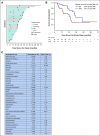
Patient outcomes (survival and safety): (A) Swimmer plot showing disease status and outcomes for all patients with a follow-up of 72 months. (B) Kaplan-Meier survival curve depicting PFS and OS for all patients. (C) AEs—treatment-related toxicity. Bars represent on-study and follow-up period. AE, adverse event; NR, not reached; OS, overall survival; PFS, progression-free survival.
FIG 3.
FIG 3.
Outcome-related gene and mutational signatures of HIPEC responders. (A) Hierarchical clustered analysis of significantly changed genes in tumor samples of 15 patients. Responders were categorized into good responders (PFS > 12 months) and poor responders (PFS < 12 months). (B) Kaplan-Meier survival curve depicting PFS of good versus poor responders. (C) Kegg pathway gene set enrichment analysis from tumors of HIPEC responders demonstrate the top significantly upregulated and downregulated pathways (FDR < 0.05). Several immune-related pathways are upregulated in good responders, most prominently TNFα signaling via NFκB, while metabolic pathways are downregulated. (D) Oncoplot of the top 20 mutated genes. The upper bar plot indicates the number of intergenic somatic variants per patient while the right bar plot shows the number of variants per gene. The CADD score is shown on the left. The mutation types, histology types, and response to HIPEC are noted below the oncoplot. CADD, combined annotation dependent depletion; FDR, false discovery rate; HIPEC, hyperthermic intraperitoneal chemotherapy; HR, hazard ratio; NES, normalized enrichment score; NFκB, nuclear factor kappa B; PFS, progression-free survival; q. val., q value; TMB, tumor mutational burden; TNFα, tumor necrosis factor α.
FIG 4.
FIG 4.
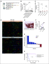
Tumor microenvironment changes induced by HIPEC. Multiplex immunofluorescence estimation of tumor-infiltrating immune subsets and PD-1 expression in matched pre- and post HIPEC tumors. (A) Pre- and post-HIPEC tumors were stained with TIL markers (CD3, CD8, FOXP3), PD-1, for the following phenotypes: CD4+ conventional T cells (CD3+ CD8– FOXP3– cells), CD4+ regulatory T cells (CD3+ CD8– FOXP3+), and CD8+ T cells (CD3+ CD8+). (B) Cell density of CD8+ T cells, CD4+ conventional T cells, and CD4+ regulatory T cells do not change with HIPEC treatment. (C) Evaluation of PD-1 expression in CD8+ T cells. PD-1 expression rises in CD8+ T cells within cancer islands (cancer), but not in stroma. (D) Representative immunofluorescence demonstrating increased PD-1 staining of CD8+ T cells within cancer islands after HIPEC while stromal PD-1 expression remained stable after HIPEC. (E) CK was used as a marker for cancer islands to delineate it from stroma. (F) PD-1 expression changes in individual patients before and after HIPEC in CD8+ T cells (stroma). The patient with the highest rise in %PD-1 was patient 1, an exceptional responder with PFS of 5 years and demonstrated the largest PD-1 increase. The only patient with decreased PD-1 expression was patient 8, who was the only patient with clear cell histology and had a poor survival (PFS < 12 months). (G) PD-1 expression changes (delta %PD-1) after HIPEC and correlation with response. %PD-1 expression changes are denoted per patient (patients 1-8). Responders were categorized into good (PFS > 24 months) and poor (PFS < 12 months). Poor responders have negative PD-1 expression changes while good responders have positive PD-1 expression changes, with an exception responder (patient 1) demonstrating the largest PD-1 rise. (H) Kaplan-Meier curves of PFS in patients with high (pink) versus low (blue) delta %PD-1 expression changes in CD8+ TILs. %PD-1 expression was dichotomized on the basis of a median delta %PD-1 threshold. CK, cytokeratin; DAPI, 4',6-diamidino-2-phenylindole; HIPEC, hyperthermic intraperitoneal chemotherapy; NS, not significant; PD-1, programmed cell death protein 1; PFS, progression-free survival; TIL, tumor-infiltrating lymphocyte.
FIG 5.
FIG 5.
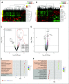
Gene and pathway changes of pre- and post-HIPEC in metastatic tumor and normal patient samples. (A and B) Hierarchical clustered analysis of (A) differentially expressed genes in metastatic tumors and (B) normal samples. Cluster 1 was enriched for pre-HIPEC; cluster 2 was enriched with post-HIPEC. (C) Volcano plot displaying gene expression post-HIPEC by log2-fold change (x-axis) and minus log10 P value (y-axis) in tumor samples. Significantly upregulated genes of ≥ 2-fold expression are in red while statistically significantly downregulated genes of ≥ 2-fold expression are in green. The top five differentially expressed genes with fold change (post- v pre-HIPEC) and P value shown for tumor samples are shown. (D) Volcano plot displaying gene expression post-HIPEC by log2-fold change (x-axis) and minus log10 P value (y-axis) in normal samples. (E and F) Kegg pathway gene set enrichment analysis of (E) HIPEC metastatic tumors and (F) normal samples demonstrating the top significantly upregulated and downregulated pathways (FDR < 0.05). Several immune-related pathways are upregulated, and DNA replication pathways are downregulated in tumor samples. Normal HIPEC samples demonstrate significant downregulation of metabolic pathways. FC, fold change; FDR, false discovery rate; HIPEC, hyperthermic intraperitoneal chemotherapy; NES, normalized enrichment score; q. val., q value; TCA, tricarboxylic acid.

References

    1. de Bree E, Tsiftsis DD: Experimental and pharmacokinetic studies in intraperitoneal chemotherapy: From laboratory bench to bedside. Recent Results Cancer Res 169:53-73, 2007 - PubMed
    1. Armstrong DK, Bundy B, Wenzel L, et al. : Intraperitoneal cisplatin and paclitaxel in ovarian cancer. N Engl J Med 354:34-43, 2006 - PubMed
    1. Mackay HJ, Kohn EC: Intraperitoneal chemotherapy: Hot, timely, and relevant? Cancer 126:5206-5209, 2020 - PubMed
    1. Dellinger TH, Han ES: State of the Science: The role of HIPEC in the treatment of ovarian cancer. Gynecol Oncol 160:364-368, 2021 - PubMed
    1. van Driel WJ, Koole SN, Sikorska K, et al. : Hyperthermic intraperitoneal chemotherapy in ovarian cancer. N Engl J Med 378:230-240, 2018 - PubMed

Publication types

MeSH terms

Associated data