. 2022 Mar 31;17(3):e0266090.
doi: 10.1371/journal.pone.0266090. eCollection 2022.

Exploring a peptide nucleic acid-based antisense approach for CD5 targeting in chronic lymphocytic leukemia

Affiliations

Exploring a peptide nucleic acid-based antisense approach for CD5 targeting in chronic lymphocytic leukemia

Elena Cesaro et al. PLoS One. .

Abstract

We herein report an innovative antisense approach based on Peptide Nucleic Acids (PNAs) to down-modulate CD5 expression levels in chronic lymphocytic leukemia (CLL). Using bioinformatics tools, we selected a 12-mer tract of the CD5 mRNA as the molecular target and synthesized the complementary and control PNA strands bearing a serine phosphate dipeptide tail to enhance their water solubility and bioavailability. The specific recognition of the 12-mer DNA strand, corresponding to the target mRNA sequence by the complementary PNA strand, was confirmed by non-denaturing polyacrylamide gel electrophoresis, thermal difference spectroscopy, circular dichroism (CD), and CD melting studies. Cytofluorimetric assays and real-time PCR analysis demonstrated the downregulation of CD5 expression due to incubation with the anti-CD5 PNA at RNA and protein levels in Jurkat cell line and peripheral blood mononuclear cells from B-CLL patients. Interestingly, we also observed that transfection with the anti-CD5 PNA increases apoptotic response induced by fludarabine in B-CLL cells. The herein reported results suggest that PNAs could represent a potential candidate for the development of antisense therapeutic agents in CLL.


Conflict of interest statement

The authors have declared that no competing interests exist.

Figures

Fig 1
Fig 1
PAGE in 100 mM PBS of: A) PNA (lane 1), DNA mixed with PNA (lane 2), DNA (lane 3), scrambled PNA (lane 4), and DNA mixed with scrambled PNA (lane 5); B) pyrimidine-rich control DNA mixed with PNA (lane 1), purine-rich control DNA mixed with PNA (lane 2), PNA (lane 3), pyrimidine-rich control DNA (lane 4), and purine-rich control DNA (lane 5). All mixtures were prepared at a 1:3 DNA/PNA ratio.
Fig 2
Fig 2
CD profile of the single-strand DNA alone (solid black line, panel A and B) and after annealing with PNA or scrambled PNA (dashed line, A and B respectively); samples were dissolved in 100 mM PBS at pH 6.8. The arithmetic sum of DNA and PNA or DNA and scrambled PNA is reported as the red line (panel A and B, respectively). The CD profile of PNA or scrambled PNA alone is reported as the dotted line (panel A and B, respectively). All spectra were acquired at 5°C in the range 200–300 nm and normalized at 300 nm; Table (C) λ values of CD minima and maxima of each sample.
Fig 3
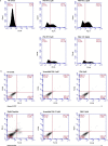
Fig 3. Evaluation of PNA transfection efficiency and cell death analysis in Jurkat cells.
(A) Flow cytometric histograms of Jurkat cells transfected with PNA-FITC to measure PNA delivery efficiency into the cells. Different concentrations of PNA-FITC were used (1, 2.5, 5, and 10 μM), and 48 h after transfection the cells were harvested and analyzed by flow cytometry. A marker was placed on non-transfected cells (NT) and FL1 fluorescence measured as percentage of cells uptaking the PNA. The figure shows two peaks on transfected cells that can be interpreted as the positive (V3-R) and negative (V3-L) datasets. (B) Flow cytometric evaluation of cell death. Two different concentrations (1.0 and 2.5 μM) of PNA or scrambled PNA were used for transfection. Necrotic and apoptotic cells were detected by annexin V and PI staining followed by flow cytometry analysis 48 h after transfection. The LR, UR, and UL quadrants measure the cells Annexin V+/PI(early apoptosis), Annexin V+/PI+ (late apoptosis), and Annexin V/PI+ (necrosis), respectively. The LL quadrants represent the percentage of double-negative cells. No variation in Annexin V and PI percentage was observed in PNA-treated cells compared to NT, or cells treated with the scrambled PNA. In contrast, the treatment with 20 μM cisplatin for 24 h used as cell death positive control, induced Jurkat cell death as expected.
Fig 4
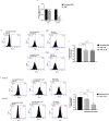
Fig 4. Evaluation of PNA effect on CD5 expression.
Jurkat cells were transfected with two different concentrations of scrambled PNA or PNA (1.0 and 2.5 μM). (A) Evaluation of CD5 mRNA expression levels was performed by RT-qPCR 48 h after transfection by comparing PNA transfected cells to cells transfected with scrambled PNA at both concentrations. Cells transfected with PNA showed a decreased CD5 mRNA expression, differently from cells transfected with scrambled PNA used as negative control. HPRT was used as a reference gene for the relative normalization of gene expression analysis. The data shown are the mean ± SD of three independent experiments. Differences were considered significant when p ≤ 0.05 (*) and p ≤ 0.0001 (**) versus scrambled PNA. (B) Flow cytometric histograms of membrane CD5 expression in Jurkat cells transfected for 48 h with PNA-FITC or the scrambled PNA-FITC. A marker was placed on the control isotype IgG stained cells and FL2 fluorescence measured as percentage of CD5 positive cells. (C) Data are shown as fold change of CD5 expression of Jurkat cells transfected with PNA-FITC at 1.0 and 2.5 μM, each of them compared to the corresponding scrambled PNA-FITC, used as control (arbitrary value = 1). Values are the mean ± SD of three independent experiments. (D) Flow cytometric histograms of membrane and intracellular CD5 expression in Jurkat cells transfected for 48 h with 2.5 μM PNA-FITC or the control scrambled PNA-FITC. A marker was placed on the respective control isotype IgG stained cells and FL2 fluorescence measured as percentage of CD5 positive cells. (E) Data are shown as fold change of membrane and intracellular CD5 expression in Jurkat cells transfected with PNA-FITC, each of them compared to the corresponding scrambled PNA-FITC, used as control (arbitrary value = 1). Values are the mean ± SD of three independent experiments. Differences were considered significant when p ≤ 0.05 (*) versus scrambled PNA.
Fig 5
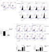
Fig 5. PNA impairs CD5 expression in B-CLL cells and sensitizes to fludarabine-induced cell death.
(A) Representative dot plots of PBMCs characterization for the expression of CD19 and CD5. (B) Flow cytometric analysis of CD5 expression in PBMCs from two B-CLL patients (P1 and P2) transfected with 1 μM scrambled PNA or PNA. A marker was placed on the relative control isotype IgG stained cells and FL4 fluorescence measured as percentage of CD5 positive cells. To evaluate CD5 reduction, for each patient the ratio between the percentage values of PNA and scrambled PNA subtracted of their corresponding IgG background (values shown in each panel) was calculated. The obtained scrambled PNA percentage was set as 100. The ratio values for P1 and P2 were 78% and 73%, respectively, corresponding to a reduction of 22% and 27% (average reduction of about 25%). (C) PNA transfection significantly decreased CD5 transcript levels compared to scrambled PNA in PBMCs from three CLL patients (P1, P3, and P5). p ≤ 0.0001 (**). (D) Representative flow cytometric histograms of Annexin V/PI staining. PBMCs from two B-CLL patients (P3 and P4) transfected with 1 μM scrambled PNA or PNA were incubated in the absence (-fludarabine, top panels) or presence (+ fludarabine, bottom panels) of 9 μM fludarabine for 72 h. (E) Histograms showing cell death values (Annexin V+/PIcells and Annexin V+/PI+ cells) obtained from PBMCs of patients P3 and P4. Data shown represent the mean ± SD from technical triplicates.

References

    1. Nabhan C, Rosen ST. Chronic Lymphocytic Leukemia: A Clinical Review. JAMA—J Am Med Assoc. 2014;312: 2265–2276. doi: 10.1001/jama.2014.14553 - DOI - PubMed
    1. Bosch F, Dalla-Favera R. Chronic lymphocytic leukaemia: from genetics to treatment. Nat Rev Clin Oncol. 2019;16: 684–701. doi: 10.1038/s41571-019-0239-8 - DOI - PubMed
    1. Starostka D, Kriegova E, Kudelka M, Mikula P, Zehnalova S, Radvansky M, et al.. Quantitative assessment of informative immunophenotypic markers increases the diagnostic value of immunophenotyping in mature CD5-positive B-cell neoplasms. Cytom Part B—Clin Cytom. 2018;94: 576–587. doi: 10.1002/cyto.b.21607 - DOI - PubMed
    1. Friedman DR, Guadalupe E, Volkheimer A, Moore JO, Weinberg JB. Clinical outcomes in chronic lymphocytic leukaemia associated with expression of CD5, a negative regulator of B-cell receptor signalling. Br J Haematol. 2018;183: 747–754. doi: 10.1111/bjh.15632 - DOI - PMC - PubMed
    1. Jaseb K, Purrahman D, Shahrabi S, Ghanavat M, Rezaeean H, Saki N. Prognostic significance of aberrant CD5 expression in B-cell leukemia. Oncol Rev. 2019;13: 77–82. doi: 10.4081/oncol.2019.400 - DOI - PMC - PubMed

Publication types

MeSH terms