. 2022 Mar 31;17(3):e0266395.
doi: 10.1371/journal.pone.0266395. eCollection 2022.

Tracing back variations in archaeal ESCRT-based cell division to protein domain architectures

Affiliations

Tracing back variations in archaeal ESCRT-based cell division to protein domain architectures

Béla P Frohn et al. PLoS One. .

Abstract

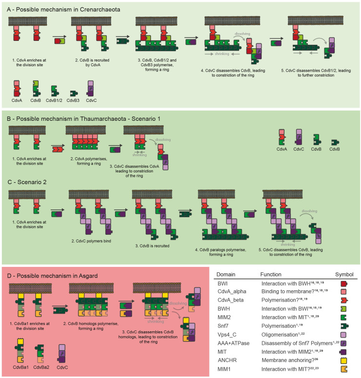
The Endosomal Sorting Complex Required for Transport (ESCRT) system is a multi-protein machinery that is involved in cell division of both Eukaryotes and Archaea. This spread across domains of life suggests that a precursor ESCRT machinery existed already at an evolutionary early stage of life, making it a promising candidate for the (re)construction of a minimal cell division machinery. There are, however, only few experimental data about ESCRT machineries in Archaea, due to high technical challenges in cultivation and microscopy. Here, we analyse the proteins of ESCRT machineries in archaea bioinformatically on a protein domain level, to enable mechanistical comparison without such challenging experiments. First, we infer that there are at least three different cell division mechanisms utilizing ESCRT proteins in archaea, probably similar in their constriction mechanisms but different in membrane tethering. Second, we show that ESCRT proteins in the archaeal super-phylum Asgard are highly similar to eukaryotic ESCRT proteins, strengthening the recently developed idea that all Eukaryotes descended from archaea. Third, we reconstruct a plausible evolutionary development of ESCRT machineries and suggest that a simple ESCRT-based constriction machinery existed in the last archaeal common ancestor. These findings not only give very interesting insights into the likely evolution of cell division in Archaea and Eukaryotes, but also offer new research avenues by suggesting hypothesis-driven experiments for both, cell biology and bottom-up synthetic biology.


Conflict of interest statement

The authors have declared that no competing interests exist.

Figures

Fig 1
Fig 1. Distribution of CdvABC homologs in archaea and their domain composition.
Existing homologs are displayed as filled circles, missing homologs as empty circles. Red circles represent CdvA homologs, green circles represent CdvB homologs and purple circles represent CdvC homologs. Where parts of the sequence of a homolog could be assigned to specific protein domains, this is visualised by segmentation of the filled circles with deeper colour. Organisms are arranged by phylogenetic relationship [25] and coloured by super-phyla. The TACK super-phylum is further divided into Thaumarchaeota and Crenarchaeota. 51 organisms are display, 37 of them possess at least one CdvABC homolog, 14 do not possess any CdvABC homologs. Abbreviations: BWI: Broken Winged-Helix Interaction Site, CdvA_alpha: Alpha-helix rich CdvA domain, CdvA_beta: Beta-sheet rich CdvA domain, BWH: Broken Winged Helix domain, MIM1 / -2: MIT-interacting-motif 1 / -2, Snf7: Vacuolar-sorting protein SNF7 domain, Vps4_C: Vps4 C terminal oligomerisation domain, MIT: Microtubule Interacting and Trafficking molecule domain, ANCHR: N-terminal membrane binding domain in ESCRT-III proteins.
Fig 2
Fig 2. Potential protein-protein interactions of archaeal CdvABC homologs.
Proteins are displayed as sets of building blocks, each building block representing one protein domain. Five potential domain-based interactions derived from literature are displayed in the top, grey numbers indicating the literature references where these domain-domain interactions were described. Based on the potential domain-domain interactions, protein-protein interaction networks are shown, where arrows indicate a potential interaction between two domains of the connected proteins. Literature about polymerisation of the CdvA_beta domain varies, so it is displayed in grey instead of black. The networks visualise the interactions of proteins that most likely existed in the last common ancestors of each of the three phylogenetic groups Crenarchaeota, Thaumarchaeota and Asgard archaea (see also Fig 6). Thus, this visualises conceptual differences between the three phylogenetic groups, and does not reflect the specific protein interactions of all organisms found today. Abbreviations and colours as in Fig 1.
Fig 3
Fig 3. Phylogenetic tree of CdvB homologs calculated via Bayesian phylogeny.
Background colours as in Fig 1 plus Eukaryotes (ESCRT-III proteins of Homo sapiens and Saccharomyces cerevisiae) in yellow, proteins schematically depicted as sets of domains as in Fig 2. In Crenarchaeota, CdvB homologs split into three phylogenetic clusters (CdvB, CdvB1/2 and CdvB3) where within each cluster most homologs have the same domain composition. In Thaumarchaeota, no clear phylogenetic clustering is visible. In Asgard archaea, CdvB homologs split into two branches, with clear differences in domain architecture between clusters. They group together with the two eukaryotic ESCRT-III groups Vps2/24/46 (CdvBa1) and Vps20/32/60 (CdvBa2), indicating a shared evolutionary history of ESCRT-III proteins in Asgard archaea and Eukaryotes. Single proteins that do not match the clusters are indicated by arrows. These might be the results of horizontal gene transfer or contamination of metagenome data. A: Two Euryarchaeota CdvB proteins (Thermoplasa acidophilum, UniProt ID Q9HIZ5 and Thermoplasma volcanium, Q97BR8). B: Thaumarchaeon Nitrosopumilus maritimus, A9A4K8. C: Fervidococcus fontis, domain architecture of a CdvB protein (Snf7 and MIM2), I0A2N3. D: Candidatus Heimdallarchaeon, A0A523XLA6.
Fig 4
Fig 4. Possible mechanisms of Cdv-based cell division in Crenarchaeota, Thaumarchaeota and Asgard.
Proteins are displayed as sets of building blocks as in Fig 2. Importantly, these mechanisms are not results of mathematical modelling, but are a qualitative explanation based on PPI networks (S1 Fig). A: The possible mechanism in Crenarchaeota is characterised by a sequential enrichment of CdvA, CdvB, CdvB1/2 and -3, followed by depolymerisation by CdvC. B: In the first scenario in Thaumarchaeota, CdvB homologs are not involved at all and ring-formation is instead relying on polymerisation of CdvA. C: The second scenario in Thaumarchaeota involves CdvB polymerisation, which is connected to CdvA by CdvC acting as linker. We strongly doubt this scenario. D: In Asgard archaea, the possible mechanism starts by CdvBa1 homologs binding to the membrane, differing from TACK archaea. CdvB homologs may then polymerise, form a ring, and get depolymerised by CdvC.
Fig 5
Fig 5. Multiple sequence alignment and potential domains of Asgard CdvB homologs.
Predicted secondary structure is indicated by background colour, amino acids matching regular expressions of specific domains as defined in the literature are indicated by font colour. Prolines in red. Protein sequences of the different clades in the phylogeny (Fig 3), CdvBa1 and CdvBa2, differ mostly at the N-terminus, where in all but one sequences of CdvBa1 an alpha helix is predicted, whilst in in all CdvBa2 proteins no alpha helix is predicted. Abbreviations: LK: Lokiarchaeum sp. GC14_75, PS: Candidatus P. syntrophicum MK-D1, OD: Candidatus Odinarchaeota archaeon LCB_4, HD: Candidatus Heimdallarchaeota archaeon, TO: Candidatus Thorarchaeota archaeon (strain OWC), TA: Candidatus Thorarchaeota archaeon strain AB_25.
Fig 6
Fig 6. Potential evolutionary development of Cdv/ESCRT machineries.
Scenario based on phylogenetic trees (Figs 2 and S4) and maximum parsimony. Domains displayed as blocks as in Figs 2–5.

References

    1. Caspi Y, Dekker C. Dividing the Archaeal Way: The Ancient Cdv Cell-Division Machinery. Front Microbiol. 2018. Mar 2;9:174. Available from: http://journal.frontiersin.org/article/10.3389/fmicb.2018.00174/full. doi: 10.3389/fmicb.2018.00174 - DOI - DOI - PMC - PubMed
    1. Makarova KS, Yutin N, Bell SD, Koonin E V. Evolution of diverse cell division and vesicle formation systems in Archaea. Nat Rev Microbiol. 2010; 8(10):731–741. doi: 10.1038/nrmicro2406 - DOI - PMC - PubMed
    1. Lindås A-C, Karlsson EA, Lindgren MT, Ettema TJG, Bernander R. A unique cell division machinery in the Archaea. Proc Natl Acad Sci U S A. 2008. Dec 2;105(48):18942–6. doi: 10.1073/pnas.0809467105 - DOI - PMC - PubMed
    1. Samson RY, Obita T, Freund SM, Williams RL, Bell SD. A role for the ESCRT system in cell division in archaea. Science 2008;322(5908):1710–1713. doi: 10.1126/science.1165322 - DOI - PMC - PubMed
    1. Pulschen AA, Mutavchiev DR, Culley S, Sebastian KN, Roubinet J, Roubinet M, et al. Live Imaging of a Hyperthermophilic Archaeon Reveals Distinct Roles for Two ESCRT-III Homologs in Ensuring a Robust and Symmetric Division. Curr Biol. 2020; 30(14):2852–2859.e4. doi: 10.1016/j.cub.2020.05.021 - DOI - PMC - PubMed

MeSH terms

Substances