. 2022 Jul 6;30(7):2554-2567.
doi: 10.1016/j.ymthe.2022.03.015. Epub 2022 Mar 28.

An SCD1-dependent mechanoresponsive pathway promotes HCC invasion and metastasis through lipid metabolic reprogramming

Affiliations

An SCD1-dependent mechanoresponsive pathway promotes HCC invasion and metastasis through lipid metabolic reprogramming

Hua-Hua Liu et al. Mol Ther. .

Abstract

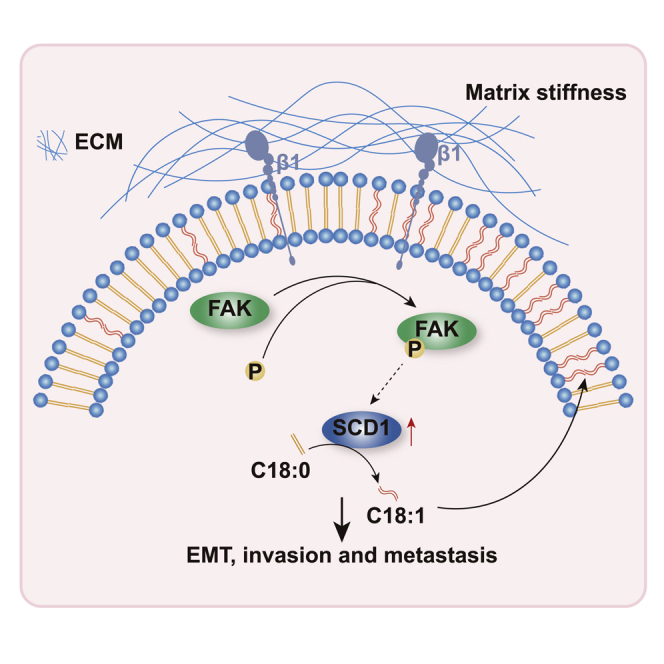
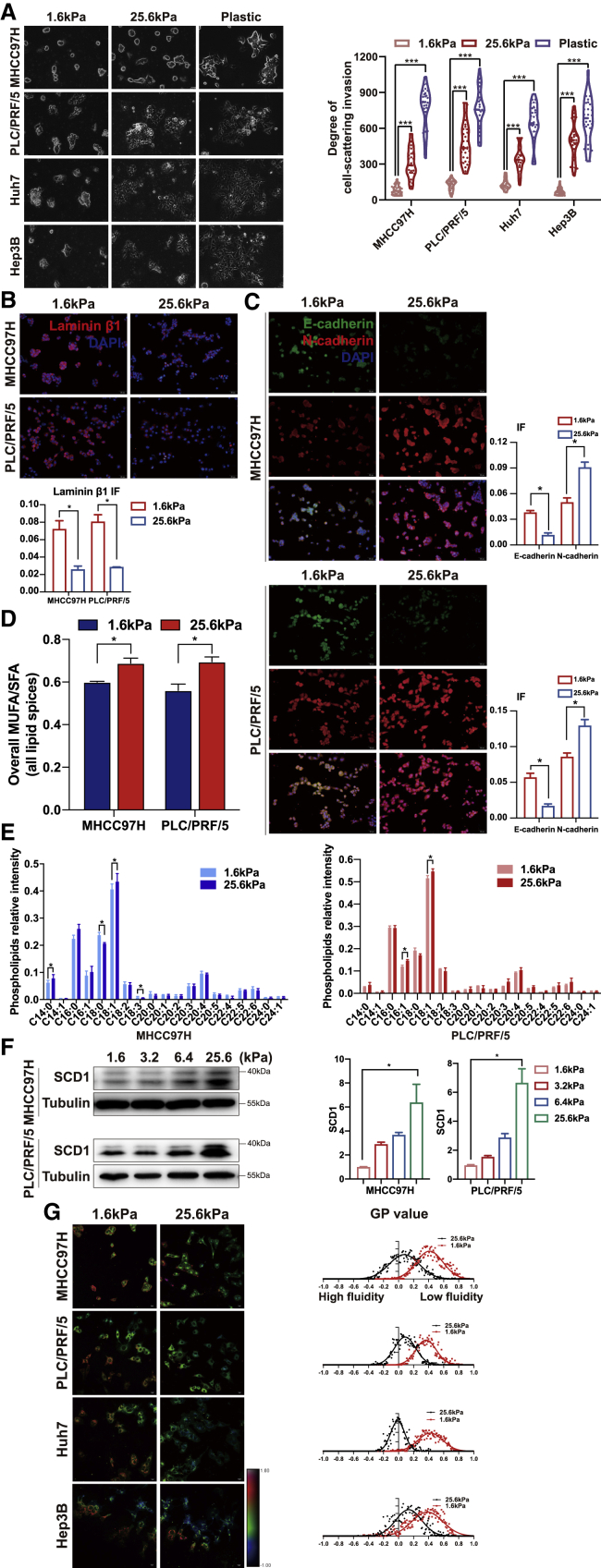
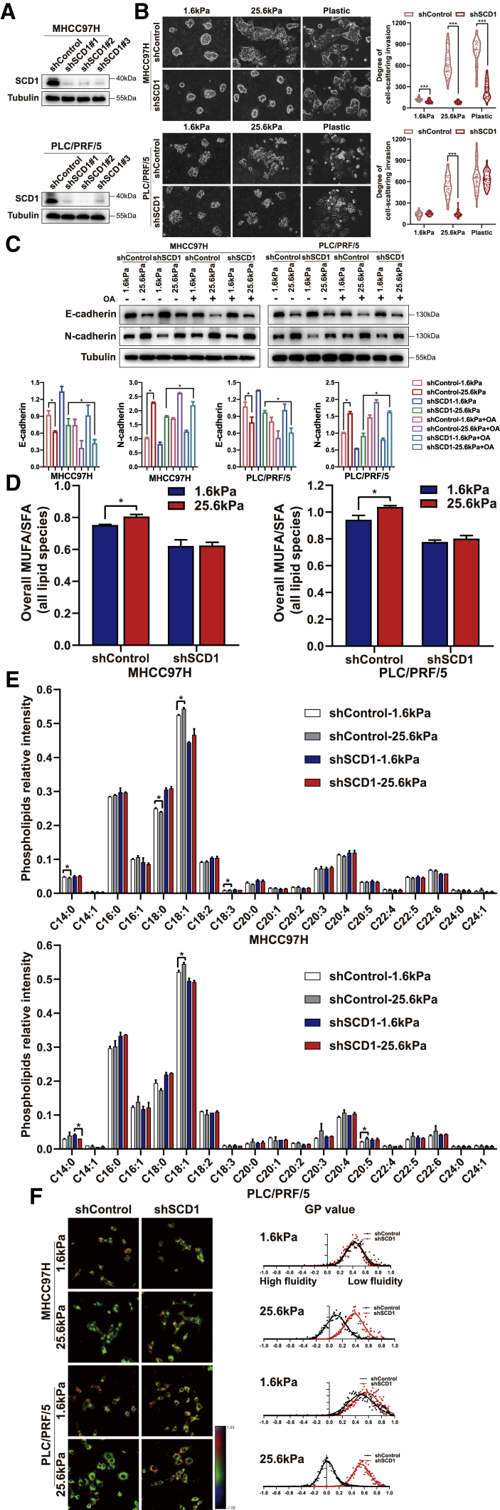
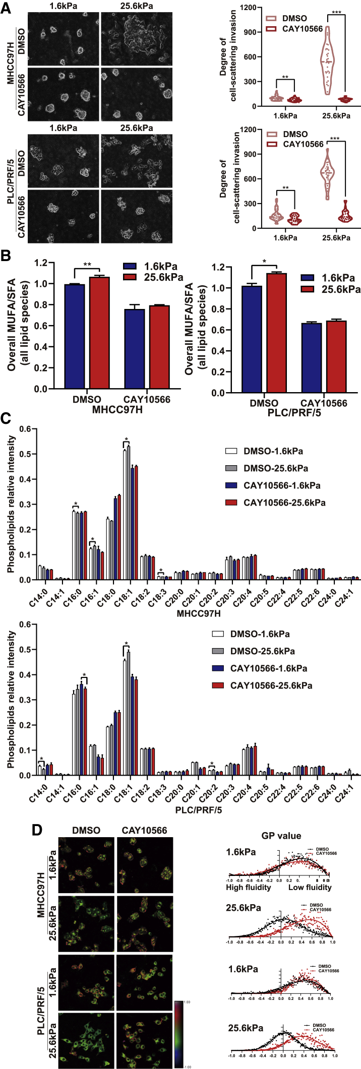
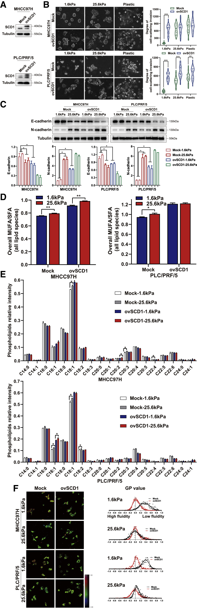
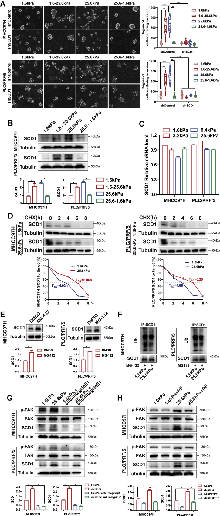
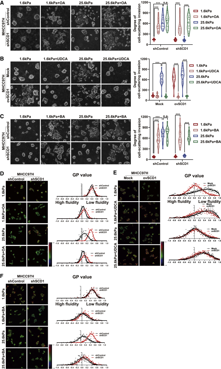
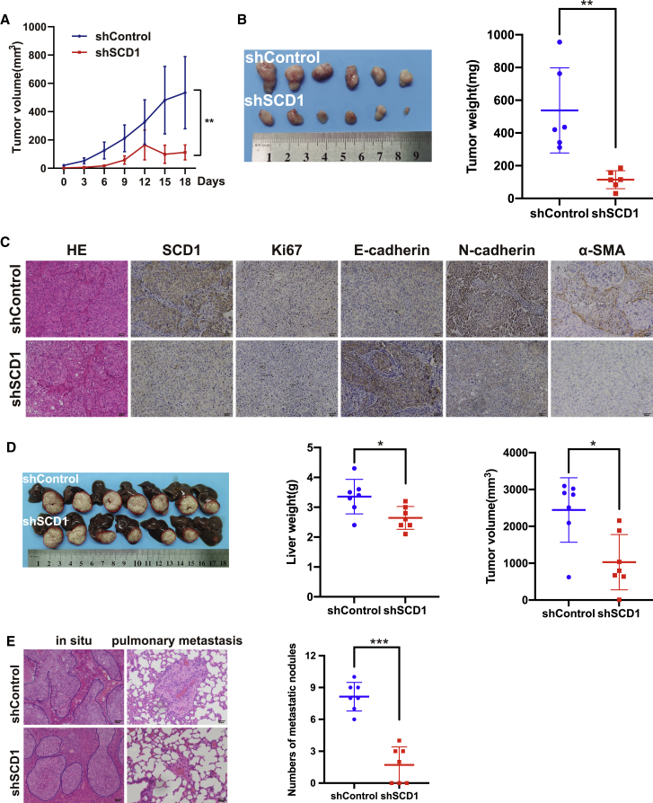
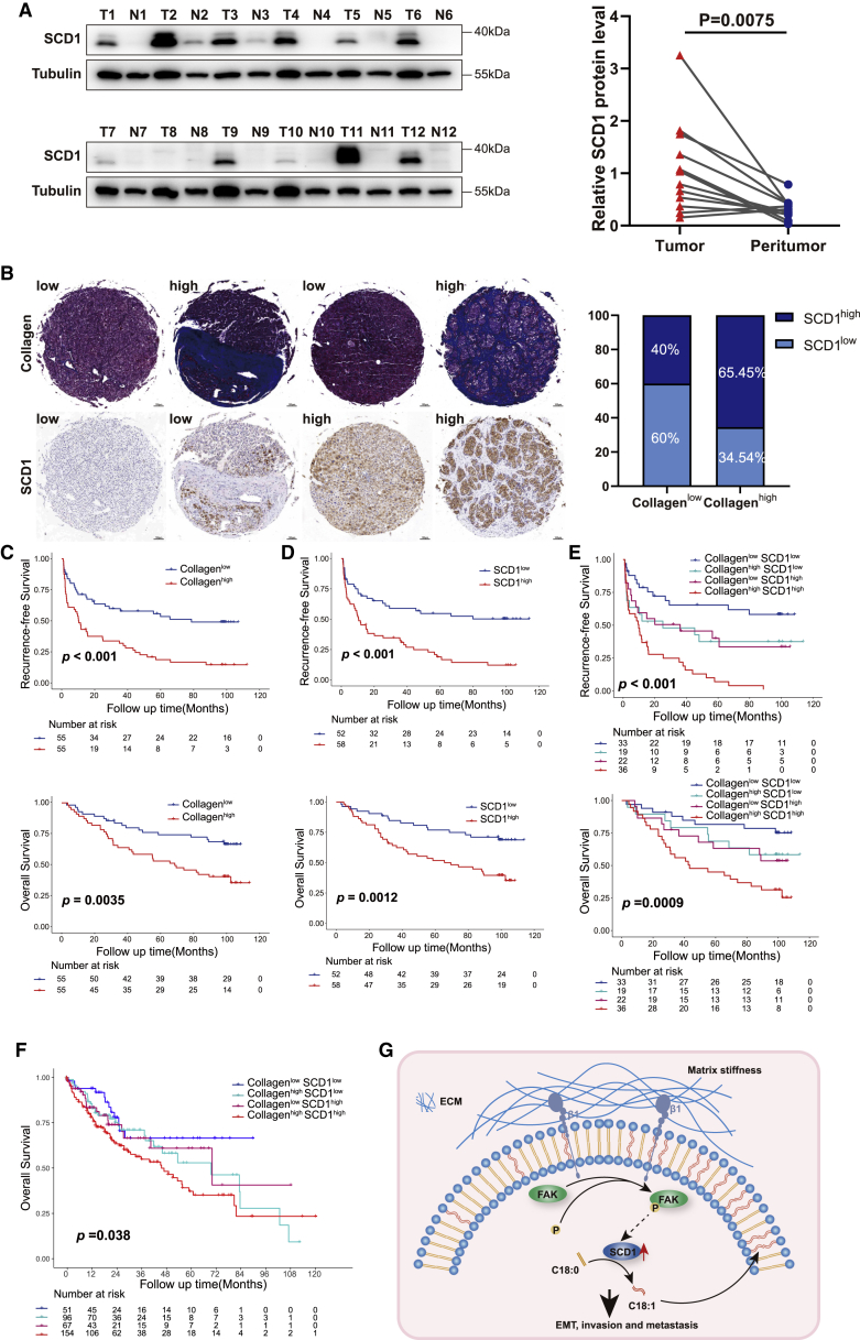
Matrix stiffness promotes hepatocellular carcinoma (HCC) metastasis. This study examined the contribution of lipid metabolic reprogramming to matrix stiffness-induced HCC metastasis. HCC cells were cultured on mechanically tunable polyacrylamide gels and subjected to lipidomic analysis. The key enzyme that responded to matrix stiffness and regulated lipid metabolism was identified. The comparative lipidomic screening revealed that stearoyl-CoA desaturase 1 (SCD1) is a mechanoresponsive enzyme that reprogrammed HCC cell lipid metabolism. The genetic and pharmacological inhibition of SCD1 expression/activity altered the cellular lipid composition, which in turn impaired plasma membrane fluidity and inhibited in vitro invasive motility of HCC cells in response to high matrix stiffness. Knockdown of SCD1 suppressed HCC invasion and metastasis in vivo. Conversely, the overexpression of SCD1 or exogenous administration of its product oleic acid augmented plasma membrane fluidity and rescued in vitro invasive migration in HCC cells cultured on soft substrates, mimicking the effects imposed by high matrix stiffness. In human HCC tissues, collagen content, a marker of increasing matrix stiffness, and increased expression of SCD1 together predicted poor survival of HCC patients. An SCD1-dependent mechanoresponsive pathway that responds to increasing matrix stiffness in the tumor microenvironment promotes HCC invasion and metastasis through lipid metabolic reprogramming.

Keywords: extracellular matrix stiffness; hepatocellular carcinoma; lipid metabolism; membrane fluidity.


Conflict of interest statement

Conflicts of interest The authors declare no competing interests.

Figures

None
Graphical abstract
Figure 1
Figure 1
Matrix stiffness promotes the invasion of HCC cells HCC cells were cultured on polyacrylamide hydrogels with the indicated rigidities (1.6 and 25.6 kPa) and plastic plates in 3D Matrigel overlay culture. (A) Bright-field images of HCC cells. The in vitro invasion of HCC cells is quantified by the degree of cell scattering using ImageJ software. (B and C) Expression of basement membrane component laminin β1 and EMT-related markers (E-cadherin and N-cadherin) was assessed by immunofluorescence staining. (D) Matrix stiffness modulated the ratio of monounsaturated/saturated fatty acids in all lipid species. (E) Phospholipids levels were determined by lipidomic mass spectrometry. (F) Western blot analyzed stiffness-induced SCD1 protein expression. (G) Hue-saturation-brightness images. The value of the minimum color (blue) was set to generalized polarization (GP) = −1 and the maximum (red) was set to 1. ∗p < 0.05, ∗∗∗p < 0.001.
Figure 2
Figure 2
SCD1 is required for matrix stiffness-induced invasion in HCC cells HCC cells expressing control (shControl) or SCD1 shRNA (shSCD1) were cultured on polyacrylamide hydrogels with the indicated rigidities (1.6 and 25.6 kPa) and plastic plates in 3D Matrigel overlay culture. (A) Efficiency of SCD1 knockdown was probed by western blot. (B) Bright-field images of HCC cells. The in vitro invasion of HCC cells is quantified by the degree of cell scattering using ImageJ software. (C) Expression of EMT-related markers E-cadherin and N-cadherin was assessed by western blot. HCC cells were treated with or without oleic acid (OA) (the product of SCD1). (D) Ratio of monounsaturated/saturated fatty acids in all lipid species. (E) Phospholipid levels were determined by lipidomic mass spectrometry. (F) Hue-saturation-brightness images of HCC cells. The value of the minimum color (blue) was set to generalized polarization (GP) = −1, and the maximum (red) was set to 1. ∗p < 0.05, ∗∗∗p < 0.001.
Figure 3
Figure 3
Pharmacological disruption of SCD1 activity suppresses matrix stiffness-induced invasion in HCC cells HCC cells were cultured on polyacrylamide hydrogels with the indicated rigidities (1.6 and 25.6 kPa) in 3D Matrigel overlay culture and treated with DMSO or SCD1 inhibitor CAY10566. (A) Bright-field images of HCC cells. The in vitro invasion of HCC cells is quantified by the degree of cell scattering using ImageJ software. (B) Ratio of monounsaturated/saturated fatty acid in all lipid species. (C) Phospholipids levels were determined by lipidomic mass spectrometry. (D) Hue-saturation-brightness images of HCC cells. The value of the minimum color (blue) was set to generalized polarization (GP) = −1, and the maximum (red) was set to 1. ∗p < 0.05, ∗∗p < 0.01, ∗∗∗p < 0.001.
Figure 4
Figure 4
Upregulation of SCD1 expression rescues the invasion of HCC cells cultured on soft matrix substrates HCC cells transfected with mock or SCD1-expressing plasmid (ovSCD1) were cultured on polyacrylamide hydrogels with the indicated rigidities (1.6 and 25.6 kPa) and plastic plates in 3D Matrigel overlay culture. (A) SCD1 protein expression was analyzed by western blot. (B) Bright-field images of HCC cells. The in vitro invasion of HCC cells was quantified by the degree of cell scattering using ImageJ software. (C) The expression of EMT-related markers (E-cadherin and N-cadherin) was assessed by western blot. (D) Ratio of monounsaturated/saturated fatty acids in all lipid species. (E) Phospholipids levels were determined by lipidomic mass spectrometry. (F) Hue-saturation-brightness images of HCC cells. The value of the minimum color (blue) was set to generalized polarization (GP) = −1 and the maximum (red) was set to 1. ∗p < 0.05, ∗∗p < 0.01, ∗∗∗p < 0.001.
Figure 5
Figure 5
Matrix stiffness modulates SCD1 expression HCC cells expressing control (shControl) or SCD1 shRNA (shSCD1) were grown on polyacrylamide hydrogels with the indicated rigidities (1.6 and 25.6 kPa) and then switched from 1.6 to 25.6 kPa (1.6–25.6 kPa) or from 25.6 to 1.6 kPa (25.6–1.6 kPa) in 3D Matrigel overlay culture (A) Representative bright-field images of HCC cells. The in vitro invasion of HCC cells is quantified by the degree of cell scattering using ImageJ software. (B) SCD1 expression was analyzed by western blot. (C) qRT-PCR analysis of SCD1 mRNA expression in HCC cells on polyacrylamide hydrogels with the indicated rigidities (1.6, 3.2, 6.4, and 25.6 kPa) in 3D Matrigel overlay culture. (D) Effect of matrix stiffness on SCD1 protein stability was analyzed by western blot in the presence of cycloheximide (CHX). The half-life (T1/2) of SCD1 was calculated. HCC cells were collected at the indicated time points after exposure to CHX, and cell lysates were subjected to immunoblotting. (E) SCD1 protein expression was increased in HCC cells treated with the proteasome inhibitor MG-132. (F) The level of ubiquitinated SCD1 was reduced in HCC cells cultured on stiff matrix. (G and H) HCC cells were treated with the anti-β1-integrin antibody or p-FAK inhibitor PF562271 (PF). SCD1 expression was analyzed by western blot. ∗∗∗p < 0.001.
Figure 6
Figure 6
Membrane fluidity mediates matrix stiffness-induced cellular invasion in HCC cells (A) MHCC97H cells expressing control (shControl) or SCD1 shRNA (shSCD1) were grown on polyacrylamide hydrogels with the indicated rigidities (1.6 and 25.6 kPa) in the presence of oleic acid (OA) in 3D Matrigel overlay culture. (B) MHCC97H cells were transfected with mock or SCD1-expressing plasmid (ovSCD1) and cultured on polyacrylamide hydrogels with the indicated rigidities (1.6 and 25.6 kPa) in 3D Matrigel overlay culture and then treated with membrane-stabilizing agent UDCA. (C) MHCC97H cells expressing control (shControl) or SCD1 shRNA (shSCD1) were cultured on polyacrylamide hydrogels with the indicated rigidities (1.6 and 25.6 kPa) in 3D Matrigel overlay culture and treated with membrane-agonist compound BA. The in vitro invasion of HCC cells was quantified by the degree of cell scattering using ImageJ software and GraphPad Prism 8. (D) Hue-saturation-brightness images of MHCC97H cells expressing shControl or shSCD1 in the presence of OA. (E) Hue-saturation-brightness images of MHCC97H cells transfected with mock or ovSCD1 treated with/without UDCA. (F) Hue-saturation-brightness images of MHCC97H cells expressing shControl or shSCD1 treated with/without compound BA. ∗∗∗p < 0.001.
Figure 7
Figure 7
Knockdown of SCD1 suppresses HCC invasion and metastasis in vivo (A) The growth curve of subcutaneous tumors derived from SCD1 knockdown or control MHCC97H cells (shSCD1 versus shControl) in nude mice. (B) Weight of subcutaneous tumors from SCD1 knockdown or control MHCC97H cells (shSCD1 versus shControl). (C) Expression of SCD1, α-SMA, Ki67, and EMT-related markers (E-cadherin and N-cadherin) was analyzed by immunohistochemistry. (D) Subcutaneous tumors were orthotopically implanted into the livers of nude mice and allowed tumor development. (E) In situ liver tumors derived from SCD1 knockdown versus shControl MHCC97H cells presented a less invasive tumor edge and a decrease in the number of distant lung metastases, as indicated by H&E staining. ∗∗p < 0.01, ∗∗∗p < 0.001.
Figure 8
Figure 8
Increased collagen content and SCD1 upregulation synergistically predict poor prognosis in HCC patients (A) SCD1 protein expression in the tumor and non-tumoral tissues detected by western blot. T, tumor tissues; N, non-tumoral tissues. (B) Representative images of HCC tissues analyzed for intratumoral collagen content by Masson’s trichrome staining and SCD1 expression by immunohistochemistry staining. Correlation analysis between intratumoral collagen content and SCD1 expression. (C and D) Kaplan-Meier curve of recurrence-free survival and OS for HCC patients stratified by intratumoral collagen content or SCD1 expression. (E) Kaplan-Meier curve of recurrence-free survival and OS for HCC patients stratified by intratumoral collagen content and SCD1 expression (collagenhigh/SCD1high versus collagenhigh/SCD1low versus collagenlow/SCD1high versus collagenlow/SCD1low tumors). (F) Kaplan-Meier curve of OS for HCC patients stratified by collagen content and SCD1 expression in The Cancer Genome Atlas-HCC cohort. (G) Schematic of the proposed mechanism that an SCD1-dependent mechanoresponsive pathway responsive to increasing matrix stiffness promotes HCC invasion and metastasis through lipid metabolic reprogramming. ECM, extracellular matrix; β1, integrin β1; FAK, focal adhesion kinase; EMT, epithelial mesenchymal transformation.

References

    1. Sung H., Ferlay J., Siegel R.L., Laversanne M., Soerjomataram I., Jemal A., et al. Global cancer statistics 2020: GLOBOCAN estimates of incidence and mortality worldwide for 36 cancers in 185 countries. CA Cancer J. Clin. 2021;71:209–249. - PubMed
    1. Ishizawa T., Hasegawa K., Aoki T., Takahashi M., Inoue Y., Sano K., et al. Neither multiple tumors nor portal hypertension are surgical contraindications for hepatocellular carcinoma. Gastroenterology. 2008;134:1908–1916. - PubMed
    1. Ye Q.H., Qin L.X., Forgues M., He P., Kim J.W., Peng A.C., et al. Predicting hepatitis B virus-positive metastatic hepatocellular carcinomas using gene expression profiling and supervised machine learning. Nat. Med. 2003;9:416–423. - PubMed
    1. Tang Z.Y., Ye S.L., Liu Y.K., Qin L.X., Sun H.C., Ye Q.H., et al. A decade's studies on metastasis of hepatocellular carcinoma. J. Cancer Res. Clin. Oncol. 2004;130:187–196. - PubMed
    1. Affo S., Yu L.X., Schwabe R.F. The role of cancer-associated fibroblasts and fibrosis in liver cancer. Annu. Rev. Pathol. 2017;12:153–186. - PMC - PubMed

Publication types