. 2022 Jun:52:102299.
doi: 10.1016/j.redox.2022.102299. Epub 2022 Mar 23.

Synthesis and cellular evaluation of click-chemistry probes to study the biological effects of alpha, beta-unsaturated carbonyls

Affiliations

Synthesis and cellular evaluation of click-chemistry probes to study the biological effects of alpha, beta-unsaturated carbonyls

Chiara Morozzi et al. Redox Biol. 2022 Jun.

Abstract

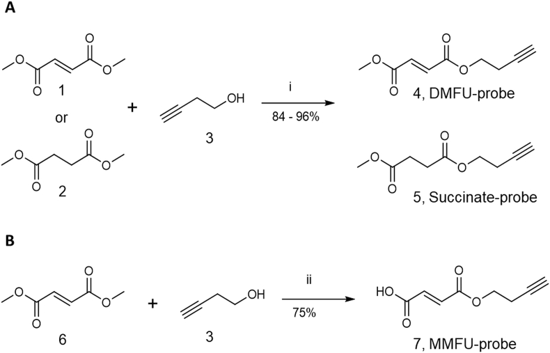
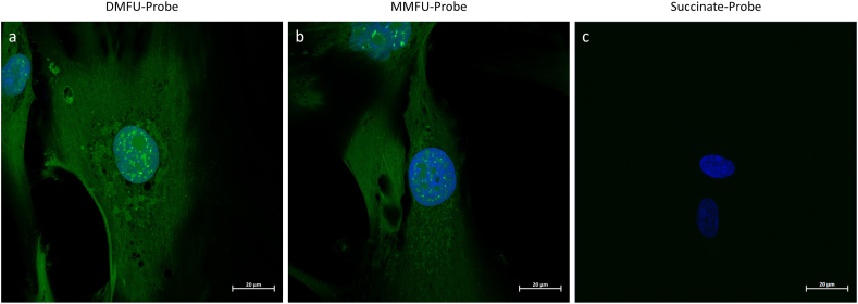
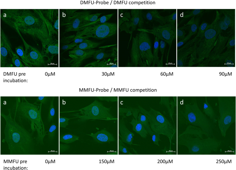
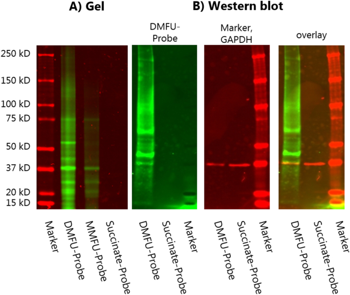
Humans are commonly exposed to α,β-unsaturated carbonyls as both environmental toxins (e.g. acrolein) and therapeutic drugs (e.g. dimethylfumarate, DMFU, a front-line drug for the treatment of multiple sclerosis and psoriasis). These compounds undergo rapid Michael addition reactions with amine, imidazole and thiol groups on biological targets, with reaction at protein Cys residues being a major reaction pathway. However, the cellular targets of these species (the 'adductome') are poorly understood due to the absence of readily identifiable tags or reporter groups (chromophores/fluorophores or antigens) on many α,β-unsaturated carbonyls. Here we report a 'proof of concept' study in which we synthesize novel α,β-unsaturated carbonyls containing an alkyne function introduced at remote sites on the α,β-unsaturated carbonyl compounds (e.g. one of the methyl groups of dimethylfumarate). The presence of this tag allows 'click-chemistry' to be used to visualize, isolate, enrich and characterize the cellular targets of such compounds. The probes show similar selectivity and reactivity to the parent compounds, and compete for cellular targets, yielding long-lived (stable) adducts that can be visualized in intact cells (such as primary human coronary artery smooth muscle cells), and extracted and enriched for subsequent target analysis. It is shown using this approach that dimethylfumarate forms adducts with multiple intracellular targets including cytoskeletal, organelle and nuclear species, with these including the rate-limiting glycolytic enzyme, glyceraldehyde-3-phosphate dehydrogenase (GAPDH). This approach should be amenable to use with multiple α,β-unsaturated carbonyls and a wide variety of targets containing nucleophilic sites.

Keywords: Click chemistry; Dimethylfumarate; Electrophile; GAPDH; Keap-1; Michael adduct; Unsaturated carbonyls.


Figures

Image 1
Graphical abstract
Fig. 1
Fig. 1
DMFU reacts with cysteine (Cys) residues on proteins via Michael addition to give S-(2-succino)cysteine adducts.
Fig. 2
Fig. 2
Summary of synthetic approaches to give (A) the DMFU (4) and succinate (5) probes, and (B) the MMFU-probe (7) using esterification with but-3-yn-1-ol (3). Reaction reagents and conditions: i) N-(3-dimethylaminopropyl)-N′-ethylcarbodiimide hydrochloride and 4-(dimethylamino)pyridine in ACN at 45 °C for 45 min. ii) N-(3-dimethylaminopropyl)-N′-ethylcarbodiimide hydrochloride and N-methylmorpholine in dry DMF, initially at 0 °C then allowed to warm up to 21 °C, overnight. For detailed descriptions of the procedures and reagents see Supplementary data.
Fig. 3
Fig. 3
Human coronary artery smooth muscle cells (HCASMC) were seeded into 24 well plates and cultured until confluent. Each well was then treated with 30 μM of DMFU (for 15 min), MMFU or succinate probe (for 4 h). After the incubation, the supernatant (500 μL) was collected, and the cells lysed in water (500 μL). Aliquots (10 μL) of each sample were then monitored over 30 min at 340 nm to quantify loss of NADH using the lactate dehydrogenase assay (see Materials and methods). Data were normalized to the untreated samples (taken as 100%) and are presented as % viability relative to the controls, as mean values +SD. No statistically-significant differences (p > 0.05) were determined between the conditions using Graphpad Prism 9 and one-way ANOVA, with testing for multiple comparisons.
Fig. 4
Fig. 4
High resolution images of human coronary artery smooth muscle cells (5 × 104 cells per well) treated with 30 μM of (a) DMFU probe (4), (b) MMFU probe (7), and (c) succinate probe (5) in growth media for 15 min (a), or 4 h (b, c) respectively. Cells were then fixed, permeabilized and then coupled to azide-derivatized Alexa 488 (3 μM) in presence of CuSO4 (2 mM) and ascorbic acid (100 mM) (see Materials and methods). Fluorescent images of the adducted species were obtained by confocal microscopy (green fluorescence, λex 488 nm, λem 543 nm). Nuclei were counter-imaged with DAPI (blue). Scale bars 20 μm. (For interpretation of the references to colour in this figure legend, the reader is referred to the Web version of this article.)
Fig. 5
Fig. 5
(a) Human coronary artery smooth muscle cells (5 × 104 cells per well) were treated with (top row) 30 μM of DMFU probe (4) or (bottom row) 30 μM of MMFU probe (7), in growth media for 15 min, then coupled to azide-derivatized Alexa 488 (3 μM) before acquisition of fluorescent images of the adducted species (green fluorescence, λex 488 nm, λem 543 nm). Nuclei were counter-imaged with DAPI (blue). (b–d) Cells were pre-incubated with (top row) 30–90 μM parent DMFU for 2 h or (bottom row) 150–200 μM of MMFU for 4 h, as a competitor for the cellular targets before reaction with the DMFU or MMFU probes. (For interpretation of the references to colour in this figure legend, the reader is referred to the Web version of this article.)
Fig. 6
Fig. 6
HCASMC were seeded onto glass cover slips and treated with DMFU-probe and MMFU-probe as described in the Materials and methods. Subsequently Alexa azide-488 was added to the cells to fluorescently-label the probe-modified proteins. Pretreatment with DMFU (left panel) or MMFU (right panel) lowered the fluorescence intensity significantly, consistent with the probes having the same specificity as the parent compounds. Pixel density of fluorescence emission from each slide was determined using an automated analysis system and a Python script (see Supplementary data) with 220000 pixels analyzed per image. Statistical analysis was carried out using Graph Pad Prism 9, using a one-way ANOVA test with post-hoc testing for multiple comparisons. Statistical significance is indicated by **** at the p < 0.001 level.
Fig. 7
Fig. 7
HCASMC were seeded in 6 well plates, cultured until confluent, then treated with 100 μM DMFU-, MMFU- or succinate-probes for 4 (DMFU-probe) or 24 h (MMFU-, succinate-probes). The cells were then subjected to ‘click chemistry’ conditions and processed as described in the Materials and methods to give fluorescently-tagged species. These were then isolated from the cultures using SP3 beads, then eluted. The tagged proteins were separated by SDS-PAGE and then imaged directly for fluorescence (panel A), or (panel B) transferred to a PVDF membrane and analyzed for the presence of the probe, immunoblotted using an anti-GAPDH primary antibody (clone GA1R) and a fluorescent secondary antibody (Azure 800). The image at the righthand side presents an overlay image of fluorescence and immunoblot data showing the co-localization of the band from GAPDH. Molecular mass markers were included as indicated.
Fig. 8
Fig. 8
HCASMC were seeded in 6 well plates, cultured until confluent, then treated with 100 μM DMFU-probe for 4 h. The cells were then subjected to ‘click chemistry’ conditions and processed as described in the Materials and methods to give fluorescently-tagged species or DAPI-stained species. The cells were then either imaged for fluorescence directly (DMFU probe and DAPI), or by using an anti-GAPDH primary antibody and a fluorescently-tagged secondary antibody as described in the Materials and methods. The top and bottom rows of images are representative data from independent experiments from a total of three. Panel A: cells probed with anti-GAPDH antibody; panel B: cells imaged directly for protein-bound DMFU-probe; panel C: cells stained with DAPI to delineate cell nuclei; panel D: composite merged image from combined panels A–C.

References

    1. Stevens J.F., Maier C.S. Acrolein: sources, metabolism, and biomolecular interactions relevant to human health and disease. Mol. Nutr. Food Res. 2008;52:7–25. - PMC - PubMed
    1. O'Brien P.J., Siraki A.G., Shangari N. Aldehyde sources, metabolism, molecular toxicity mechanisms, and possible effects on human health. Crit. Rev. Toxicol. 2005;35:609–662. - PubMed
    1. Burcham P.C. Acrolein and human disease: untangling the knotty exposure scenarios accompanying several diverse disorders. Chem. Res. Toxicol. 2017;30:145–161. - PubMed
    1. Randall M.J., Spiess P.C., Hristova M., Hondal R.J., van der Vliet A. Acrolein-induced activation of mitogen-activated protein kinase signaling is mediated by alkylation of thioredoxin reductase and thioredoxin 1. Redox Biol. 2013;1:265–275. - PMC - PubMed
    1. Poganik J.R., Aye Y. Electrophile signaling and emerging immuno- and neuro-modulatory electrophilic pharmaceuticals. Front. Aging Neurosci. 2020;12:1. - PMC - PubMed

Publication types