. 2022 Aug;12(8):1881-1894.
doi: 10.1007/s13346-022-01121-w. Epub 2022 Mar 31.

Synthesis and evaluation of wound healing properties of hydro-diab hydrogel loaded with green-synthetized AGNPS: in vitro and in ex vivo studies

Affiliations

Synthesis and evaluation of wound healing properties of hydro-diab hydrogel loaded with green-synthetized AGNPS: in vitro and in ex vivo studies

Mariarosa Ruffo et al. Drug Deliv Transl Res. 2022 Aug.

Abstract

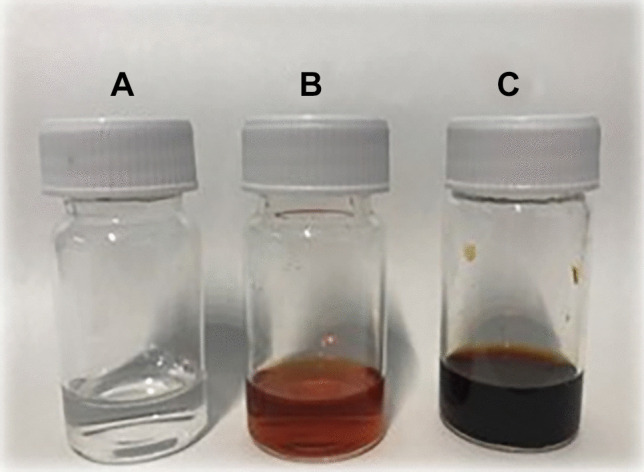
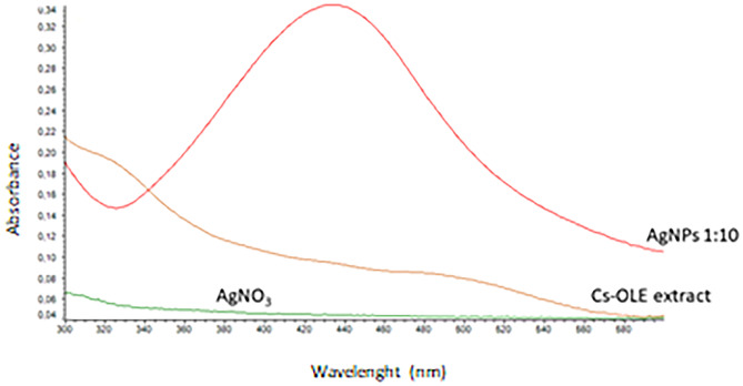
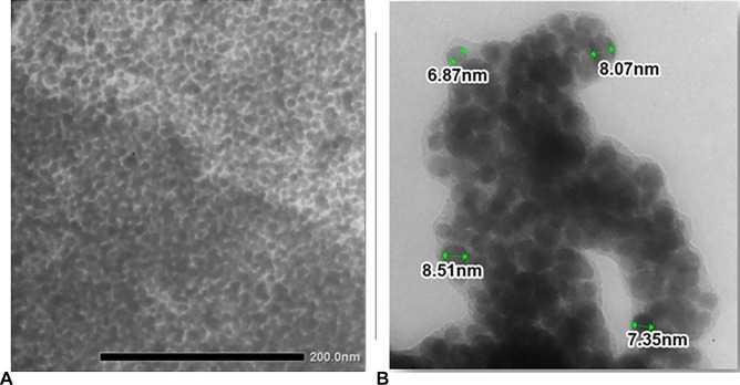
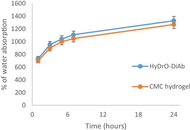
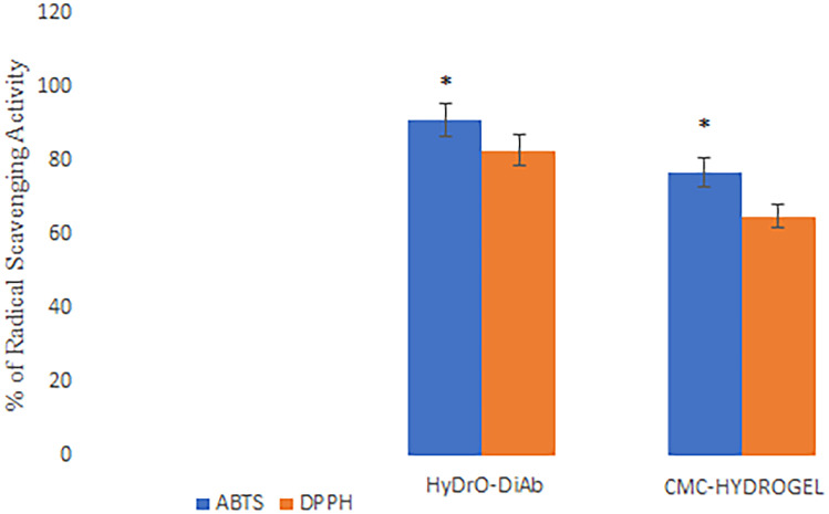
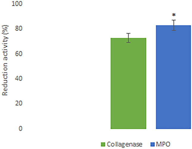
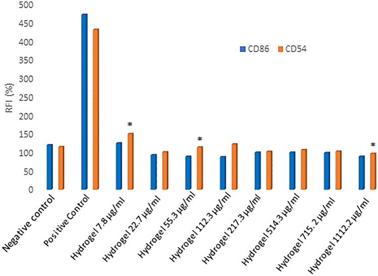
In diabetic patients, the presence of neuropathy, peripheral vascular diseases and ischemia, leads to the formation of foot ulcerations with a higher risk of infection because the normal response to bacterial infection is missing. In the aim to control and treat diabetic foot ulcerations (DFUs), wound dressings that are able to absorb exudate, to prevent infections, and to promote wound healing are needed. For this reason, the aim of the present research was to synthetize a biocompatible hydrogel (called HyDrO-DiAb) composed of carboxymethylcellulose loaded with silver nanoparticles (AgNPs) for the treatment of diabetic foot ulcers. In this study, AgNPs were obtained by a green synthesis and, then, were dissolved in a CMC hydrogel that, after a freeze drying process, becomes a flexible and porous structure. The in vitro and in ex vivo wound healing activity of the obtained HyDrO-DiAb hydrogel was evaluated.

Keywords: Diabetic foot ulcerations (DFUs); Green-synthesis; Hydrogel; Silver nanoparticles; Wound healing.


Conflict of interest statement

Not applicable.

Figures

Fig. 1
Fig. 1
AgNO3 0.01 M (A), Cs-OLE extract (B), AgNPs (C)
Fig. 2
Fig. 2
UV–vis absorption spectra of AgNO3, Cs-OLE extract and synthetized AgNPs (diluted in a ratio 1:10 with distilled water)
Fig. 3
Fig. 3
Morphologies of AgNPs (A) and dimension observed in TEM images (B)
Fig. 4
Fig. 4
Water absorption capacity of HyDrO-DiAb and CMC hydrogel. All the results are reported as mean values ± SD (n = 3)
Fig. 5
Fig. 5
Water retention capacity of HyDrO-DiAb and CMC hydrogel. All the results are reported as mean values ± SD (n = 3)
Fig. 6
Fig. 6
In vitro study release of AgNPs from hydrogel. All the results are reported as mean values ± SD (n = 3)
Fig. 7
Fig. 7
Viability of 3T3 fibroblasts cells after 24 h of incubation at 37 °C with HyDrO-DiAb. All the results are reported as mean values ± SD (n = 3)
Fig. 8
Fig. 8
Wound healing activity of HyDrO-DiAb on 3T3 fibroblast cells by scratch method (A) and evaluated after 24 h (B)
Fig. 9
Fig. 9
ABTS and DPPH radical scavenging activity of HyDrO-DiAb and CMC-hydrogel. All the results are reported as mean values ± SD (n = 3). p values < 0.05 were considered statistically significant
Fig. 10
Fig. 10
In ex vivo ability of HyDrO-DiAb to inhibit collagenase and MPO activity. All the results are reported as mean values ± SD (n = 3). p values < 0.05 were considered statistically significant
Fig. 11
Fig. 11
Expression of CD86 and CD54 after treatment with negative control, positive control and HyDrO-DiAb. Different concentrations of HyDrO-DiAb were tested to evaluate if it is a skin sensitizer. All results are reported as mean values ± SD (n = 3). p values < 0.05 were considered statistically significant

References

    1. Boyko E, Ahroni J, Cohen V, Nelson K, Heagerty P. Prediction of diabetic foot ulcer occurrence using commonly available clinical information: the Seattle diabetic foot study. Diabetes Care. 2006;29:1202–1207. doi: 10.2337/dc05-2031. - DOI - PubMed
    1. Boulton AJM, Vileikyte L, Ragnarson-Tennvall G, Apelqvist J. The global burden of diabetic foot disease. Lancet. 2005;366:1719–1724. doi: 10.1016/S0140-6736(05)67698-2. - DOI - PubMed
    1. Falanga V. Wound healing and its impairment in the diabetic foot. Lancet. 2005;366:1736–1743. doi: 10.1016/S0140-6736(05)67700-8. - DOI - PubMed
    1. Galkowska H, Wojewodzka U, Olszewski WL. Low recruitment of immune cells with increased expression of endothelial adhesion molecules in margins of the chronic diabetic foot ulcers. Wound Repair Regen. 2005;13:248–254. doi: 10.1111/j.1067-1927.2005.130306.x. - DOI - PubMed
    1. Harding KG, Jones V, Price P. Topical treatment: which dressing to choose. Diabetes Metab Res Rev. 2000;16(Suppl 1):S47–50. doi: 10.1002/1520-7560(200009/10)16:1+<::AID-DMRR133>3.0.CO;2-Q. - DOI - PubMed

Publication types