. 2022 Mar 10:10:815205.
doi: 10.3389/fcell.2022.815205. eCollection 2022.

Potential of Colostrum-Derived Exosomes for Promoting Hair Regeneration Through the Transition From Telogen to Anagen Phase

Affiliations

Potential of Colostrum-Derived Exosomes for Promoting Hair Regeneration Through the Transition From Telogen to Anagen Phase

Hyosuk Kim et al. Front Cell Dev Biol. .

Abstract

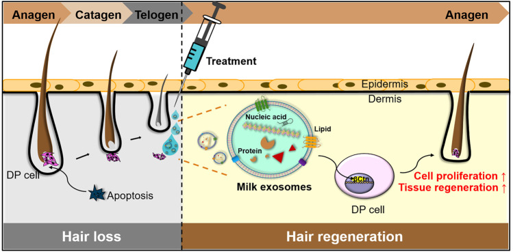
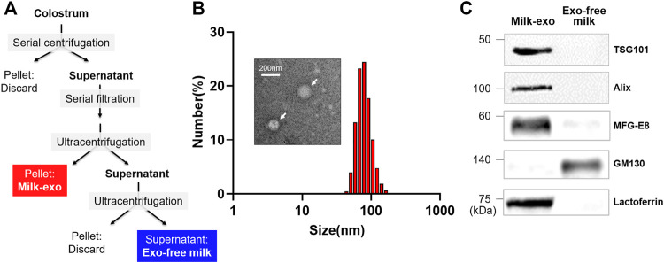
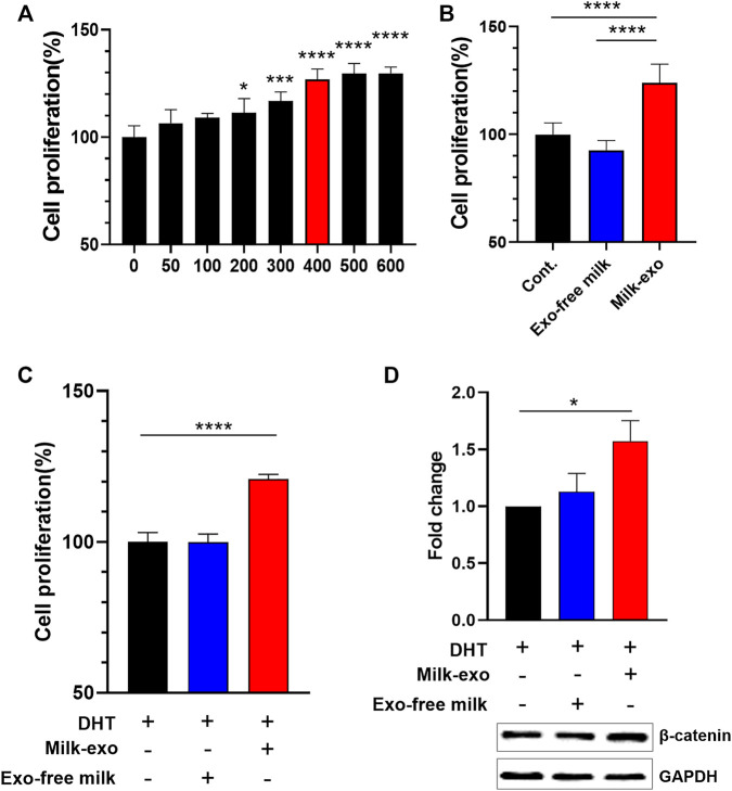
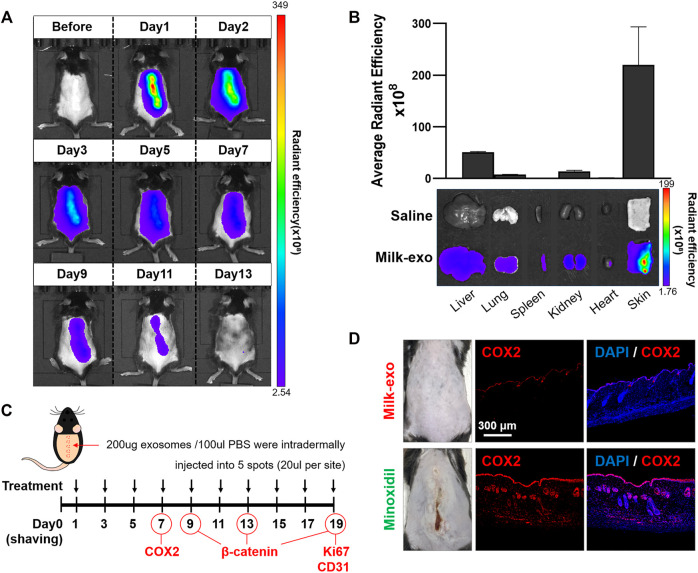
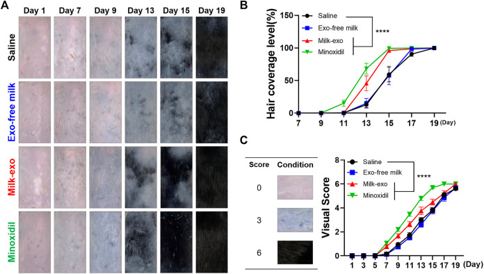
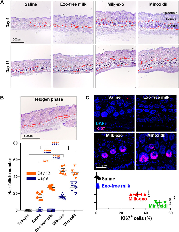
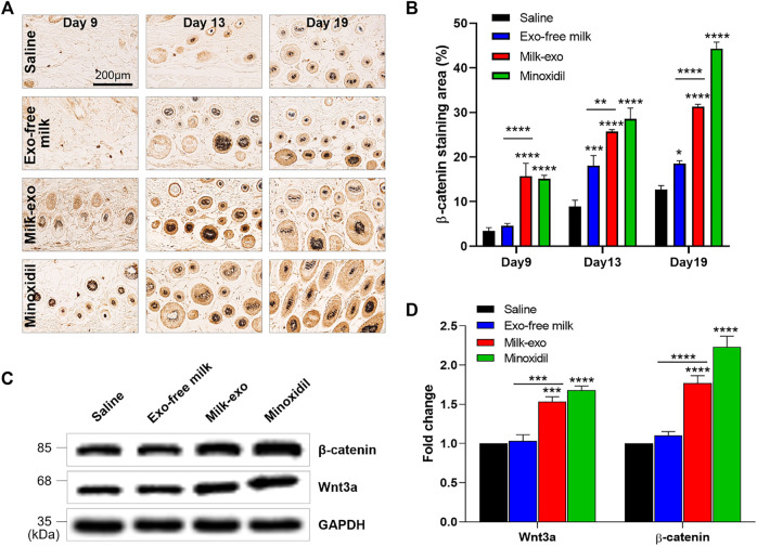
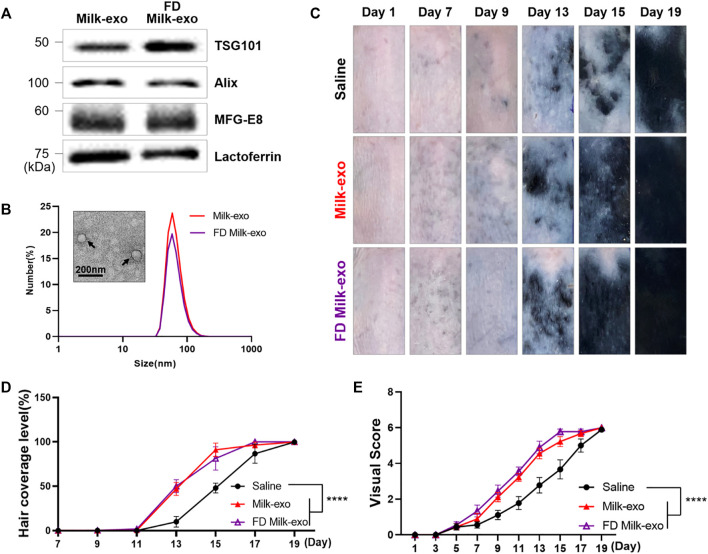
Human hair dermal papillary (DP) cells comprising mesenchymal stem cells in hair follicles contribute critically to hair growth and cycle regulation. The transition of hair follicles from telogen to anagen phase is the key to regulating hair growth, which relies heavily on the activation of DP cells. In this paper, we suggested exosomes derived from bovine colostrum (milk exosomes, Milk-exo) as a new effective non-surgical therapy for hair loss. Results showed that Milk-exo promoted the proliferation of hair DP cells and rescued dihydrotestosterone (DHT, androgen hormones)-induced arrest of follicle development. Milk-exo also induced dorsal hair re-growth in mice at the level comparable to minoxidil treatment, without associated adverse effects such as skin rashes. Our data demonstrated that Milk-exo accelerated the hair cycle transition from telogen to anagen phase by activating the Wnt/β-catenin pathway. Interestingly, Milk-exo has been found to stably retain its original properties and efficacy for hair regeneration after freeze-drying and resuspension, which is considered critical to use it as a raw material applied in different types of alopecia medicines and treatments. Overall, this study highlights a great potential of an exosome from colostrum as a therapeutic modality for hair loss.

Keywords: colostrum; dermal papilla; exosome; hair growth; lactoferrin.


Conflict of interest statement

The authors declare that the research was conducted in the absence of any commercial or financial relationships that could be construed as a potential conflict of interest.

Figures

FIGURE 1
FIGURE 1
Schematic illustration of Milk-exo-mediated phase transition from telogen to anagen for hair regeneration.
FIGURE 2
FIGURE 2
Preparation and characterization of colostrum-derived exosome (Milk-exo). (A) Scheme of the preparation procedure of Milk-exo and Exo-free milk. (B) Size distribution diagram and representative TEM image of Milk-exo. (C) Western blot analysis of common exosome markers (Tsg101 and Alix) and milk specific proteins (MFG-E8 and lactoferrin). GM130 was used as a negative control.
FIGURE 3
FIGURE 3
Effect of Milk-exo on in vitro proliferation and β-catenin expression. (A) Proliferation analysis of DP cells in response to varying concentration of Milk-exo for 24 h (n = 6). (B) Proliferation analysis of DP cells treated with Milk-exo and Exo-free milk at a concentration of 400 μg/ml for 24 h (n = 6). (C) Analysis of DP cell proliferation pattern after dihydrotestosterone (DHT) treatment (n = 6). (D) Western blots on β-catenin and GAPDH in DP cells after Milk-exo and Exo-free milk treatment for 24 h (n = 3). All data are presented as mean ± SEM (*p < 0.05, ***p < 0.001, and ****p < 0.0001 versus control).
FIGURE 4
FIGURE 4
In vivo biodistribution of Milk-exo and expression of COX2 in skin tissue. (A) Real-time in vivo imaging of Cy5.5-NHS labeled exosome in C57BL/6 mice for 13 days after intradermal injection. The mice were analyzed at the indicated times after intradermal injection of 200 µg per 100 µL of exosomes. (B) Ex vivo imaging and Milk-exo quantification of the skin and major organs on day 2 after intradermal injection of labeled exosomes (n = 3). (C) Schedule of Milk-exo treatment (black arrows) and the date of histological analysis (COX2, β-catenin, Ki67 and CD31). (D) Representative immunostaining images of the expression of COX2 in mice skin tissues treated with Milk-exo and Minoxidil. C57BL/6 mice were sacrificed on day 7 after intradermal injection every 2 days. The nuclei (blue) were stained with DAPI, and Alexa Fluor® 647 conjugated secondary antibodies (red) were used for visualization of COX2.
FIGURE 5
FIGURE 5
Hair regeneration effect of milk exosomes in C57BL/6 mice. (A) Representative images of hair regeneration after intradermal injection of saline, Exo-free milk, Milk-exo (200 μg per 100 μL), and topical application of 2.5% minoxidil. Each reagent was administrated every other day for 3 weeks. (B) Quantification of hair coverage level (n = 8, ****p < 0.0001 versus saline, two-way ANOVA, Dunnett’s multiple comparisons test). (C) Mean visual score measurements (n = 8; ****p < 0.0001, two-way ANOVA, Dunnett’s multiple comparisons test).
FIGURE 6
FIGURE 6
Histological investigation of the acceleration of telogen-to-anagen transition by milk exosomes. (A) Representative hematoxylin-eosin (H&E) images of the dorsal skin section for each group on days 9 and 13. (B) Representative image of telogen phase at day 1 after shaving and quantification of hair follicle for each groups from three tissue samples (n = 6). (C) Representative images and quantification graphs showing the expression of Ki67 on day 19 after different treatments from three tissue samples (n = 3). All data are presented as mean ± SEM (**p < 0.01, ***p < 0.001 and ****p < 0.0001).
FIGURE 7
FIGURE 7
Milk-exo activates Wnt signaling for the transition to the growth phase. (A) Immunohistochemical analysis of the expression of β-catenin in hair follicles section (day 9, day 13 and day 19). (B) Quantification of relative expression of β-catenin from five tissue samples (n = 4). Data are presented as mean ± SEM (*p < 0.05, **p < 0.01, ***p < 0.001 and ****p < 0.0001). (C) Western blot analyzing β-catenin, Wnt3a and GAPDH protein content in the dorsal skin on day 9. (D) Quantification of western blot protein levels by group. Data are presented as mean ± SEM (n = 3; ***p < 0.001 and ****p < 0.0001).
FIGURE 8
FIGURE 8
Characterization of lyophilized Milk-exo and conservation of in vivo hair growth ability. (A) Western blot analysis of exosome markers (TSG101, Alix) and milk specific proteins (MFG-E8 and lactoferrin). (B) Representative TEM image and size distribution diagram of lyophilized Milk-exo. (C) Representative images of hair regeneration after intradermal injection of saline, Milk-exo and FD Milk-exo. (D) Quantification of hair coverage level (n = 3, ****p < 0.0001 versus saline, two-way ANOVA, Dunnett’s multiple comparisons test). (E) Mean visual score measurements (n = 3; ****p < 0.0001, two-way ANOVA, Dunnett’s multiple comparisons test).

References

    1. Bai X., Lei M., Shi J., Yu Y., Qiu W., Lai X., et al. (2015). Roles of GasderminA3 in Catagen-Telogen Transition during Hair Cycling. J. Invest. Dermatol. 135, 2162–2172. 10.1038/jid.2015.147 - DOI - PMC - PubMed
    1. Bejaoui M., Villareal M. O., Isoda H. (2019). β-catenin-mediated Hair Growth Induction Effect of 3,4,5-Tri-O-Caffeoylquinic Acid. Aging 11, 4216–4237. 10.18632/aging.102048 - DOI - PMC - PubMed
    1. Botchkarev V. A., Kishimoto J. (2003). Molecular Control of Epithelial-Mesenchymal Interactions during Hair Follicle Cycling. J. Invest. Dermatol. Symp. Proc. 8, 46–55. 10.1046/j.1523-1747.2003.12171.x - DOI - PubMed
    1. Hu S., Li Z., Lutz H., Huang K., Su T., Cores J., et al. (2020). Dermal Exosomes Containing miR-218-5p Promote Hair Regeneration by Regulating β-catenin Signaling. Sci. Adv. 6, eaba1685. 10.1126/sciadv.aba1685 - DOI - PMC - PubMed
    1. Huang H.-C., Lin H., Huang M.-C. (2019). Lactoferrin Promotes Hair Growth in Mice and Increases Dermal Papilla Cell Proliferation through Erk/Akt and Wnt Signaling Pathways. Arch. Dermatol. Res. 311, 411–420. 10.1007/s00403-019-01920-1 - DOI - PMC - PubMed

LinkOut - more resources