Review
. 2022 Mar 10:9:779360.
doi: 10.3389/fchem.2021.779360. eCollection 2021.

Functional Nano-Objects by Electrostatic Self-Assembly: Structure, Switching, and Photocatalysis

Affiliations
Review

Functional Nano-Objects by Electrostatic Self-Assembly: Structure, Switching, and Photocatalysis

Anja Krieger et al. Front Chem. .

Abstract

The design of functional nano-objects by electrostatic self-assembly in solution signifies an emerging field with great potential. More specifically, the targeted combination of electrostatic interaction with other effects and interactions, such as the positioning of charges on stiff building blocks, the use of additional amphiphilic, π-π stacking building blocks, or polyelectrolytes with certain architectures, have recently promulgated electrostatic self-assembly to a principle for versatile defined structure formation. A large variety of architectures from spheres over rods and hollow spheres to networks in the size range of a few tenths to a few hundred nanometers can be formed. This review discusses the state-of-the-art of different approaches of nano-object formation by electrostatic self-assembly against the backdrop of corresponding solid materials and assemblies formed by other non-covalent interactions. In this regard, particularly promising is the facile formation of triggerable structures, i.e. size and shape switching through light, as well as the use of electrostatically assembled nano-objects for improved photocatalysis and the possible solar energy conversion in the future. Lately, this new field is eliciting an increasing amount of understanding; insights and limitations thereof are addressed in this article. Special emphasis is placed on the interconnection of molecular building block structures and the resulting nanoscale architecture via the key of thermodynamics.

Keywords: nanostructures; organic-inorganic hybrids; photocatalysis; self-assembly; stimuli-responsiveness; structure analysis; supramolecular chemistry; thermodynamics.


Conflict of interest statement

The authors declare that the research was conducted in the absence of any commercial or financial relationships that could be construed as a potential conflict of interest.

Figures

FIGURE 1
FIGURE 1
Polyelectrolyte multilayers: (A) Scheme of the film deposition process using slides and beakers. Steps 1 and 3 represent the adsorption of a polyanion and polycation, respectively, and steps 2 and 4 are washing steps for the basic buildup sequence (A/B)n. (B) Capsule formation: Schematic illustration of the polyelectrolyte deposition onto a particle process and of subsequent core decomposition; (C) SEM of nine-layer [(poly (styrene sulfonate)/poly (allylamine hydrochloride)4/poly (styrene sulfonate)] capsules. Drying, together with the topological constraints of the closed surface, results in a completely folded upper hemisphere (Donath et al., 1998). (D,E) Poly (styrene sulfonate)/poly (N, N-dimethylaminoethyl methacrylate) capsules with encapsulation of fluorescein isothiocyanate: (D) at pH = 9 and (E) release at pH = 7; Scale bar 5 μm (Xu et al., 2014). Reprinted with permission. Copyright © 1998 WILEY-VCH Verlag GmbH, Weinheim, Fed. Rep. of Germany. Copyright © 2014 American Chemical Society.
FIGURE 2
FIGURE 2
Cationic-anionic di-porphyrin needles: (A–C) TEM images of needles formed by an assembly of anionic tetra-(4-sulfonatophenyl)porphyrin and cationic tetra-(N-methyl-4-pyridyl)porphyrin with different ratios (Düring et al., 2018). Reprinted with permission. Copyright © 2018, Springer-Verlag GmbH Germany, part of Springer Nature.
FIGURE 3
FIGURE 3
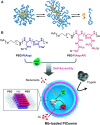
Electrostatic self-assembly with double-hydrophilic block-polyelectrolytes: (A) Formation of spherical complex coacervate core micelles from anionic poly (acrylic acid) (red) and block copolymer consisting of a cationic poly (N-methyl-2-vinylpyridinium) (blue) and a neutral poly (ethylene oxide) block (green); An increase in the salt concentration leads to the formation of an elongated worm-like structure; an even further increase in the salt concentration above the critical salt concentration leads to the separation of the polymers (van der Kooij et al., 2012); (B) Reversible myoglobin (Mb) oxygenation inside the polyion complex membrane self-assembled from a pair of oppositely charged block polyelectrolytes (Kishimura et al., 2007). Reprinted with permission. Copyright © 2007 WILEY-VCH Verlag GmbH and Co. KGaA, Weinheim.
FIGURE 4
FIGURE 4
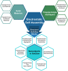
Electrostatic self-assembly: overview of established and emerging strategies.
FIGURE 5
FIGURE 5
Electrostatic self-assembly in solution: polyelectrolytes interact with oppositely charged stiff, multivalent counterions to form nano-objects with a defined size and shape; for example, cationic 5 nm-sized G4 PAMAM dendrimers interacts with dianionic dye molecules to form a 100 nm scale elongated rod-like stricture with layered internal structure.
FIGURE 6
FIGURE 6
Small-angle scattering characterization of dendrimer-naphthalene dicarboxylic acid dye assemblies: (A,B) with 1,4-naphthalene dicarboxylic acid; (C,D) with 2,3 -naphthalene dicarboxylic acid; both with a loading ratio of 2:1; left: scattering curves I(q), right: pair distance distribution functions P(r). I(q) and P(r) are in arbitrary units. 1,4-NDC yields cylindrical aggregates but 2,3-NDC spherical aggregates (Gröhn et al., 2008). Reprinted with permission. Copyright © 2008 WILEY-VCH Verlag GmbH and Co. KGaA, Weinheim.
FIGURE 7
FIGURE 7
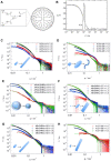
Scattering analysis of a dendrimer dye model system for electrostatic self-assembly in solution: (A) Dendrimer structure; (B) Dynamic light scattering (DLS): electric field autocorrelation function and decay time distribution at a scattering angle θ = 90° for Ar44-G4 dendrimer with a ratio l = 2.1 (Willerich et al., 2009); (C–H) SANS and resulting nanoparticle shapes as a function of dye type, dendrimer generation and loading ratio: SANS results for (C) Ar26 + G4, (D) Ar26 + G8, (E) APhAcOHRAc + G4, (F) ABnOHRAc + G7, (G) ABnOHRAc + G8, and (H) SuACAc + G8, each at varying loading ratio. Continuous lines represent the best structural fit (Mariani et al., 2016a). Reprinted with permission. Copyright © 2008 and 2015 American Chemical Society.
FIGURE 8
FIGURE 8
Shape variety in electrostatic self-assembly with different building blocks: (A) Wormlike bottle-brush polyelectrolytes and tetravalent porphyrin counterions form finite-size networks; AFM of brush and brush-porphyrin aggregates spin-coated on mica (Ruthard et al., 2009); (B) POM–dendrimer assemblies; left: static light scattering and SANS of POM–dendrimer assemblies with l = 0.7; filled symbols: SLS data, open symbols: SANS data, black line at high q: flexible cylinder fit; right: TEM image (Kutz et al., 2018); (C) Surfactant micelles connected by Ar26 ions as linkers; left: DLS, electric field autocorrelation function g1(τ) and distribution of relaxation times A(τ) for C12TAB-Ar26 assemblies; right: overview of structures formed: associated a) and individual spherical surfactant micelles b) with Ar26 molecules acting as connectors and condensed counterions, respectively, and cylindrical surfactant–dye aggregates from cylindrical surfactant micellization with condensed mutually π–π interacting Ar26 counterions c) (Kutz et al., 2016). Reprinted with permission. Copyright © 2009 American Chemical Society and © The Royal Society of Chemistry 2018 and © 2015, Springer-Verlag Berlin Heidelberg.
FIGURE 9
FIGURE 9
Shape variety achieved by electrostatic self-assembly: networks, decorated wires, stacked micelles, vesicles, tubes, ellipsoids, helices, pomegranate-like structures, discs, onion-like structures, raspberry-like structures, spheres, and rods.
FIGURE 10
FIGURE 10
Charge ratio-dependent assembly-formation in a cationic dendrimer-anionic dye system: (A) Scheme: small spheres represent individual dendrimers, cylinders larger assemblies, and red rectangles dye molecules. Bound dye molecules are not sketched for simplicity; “+” and “−” represent the sign and relative magnitude of the net charge; (B) ζ-potential in dependence on charge ratio (Willerich et al., 2010). Reprinted with permission. Copyright © 2010 American Chemical Society.
FIGURE 11
FIGURE 11
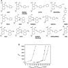
Thermodynamic study of a dendrimer-dye model system for electrostatic self-assembly in solution, elucidating the nano-objects’ size control: (A) Structural formulae of structurally related divalent and trivalent azo dyes; (B) Dendrimer aggregation number N Den in dependence on the dendrimer-dye interaction free energy ΔG dendrimer-dye (squares) and dendrimer-dye interaction enthalpy ΔH dendrimer-dye (open circles) and dye-dye interaction free energy ΔG dye_dye (triangles) for divalent dyes (Willerich and Gröhn, 2011a). Reprinted with permission. Copyright © 2011 American Chemical Society.
FIGURE 12
FIGURE 12
Elucidating the nanoscale shape control in electrostatic self-assembly by an experimental thermodynamics study: (A) Enthalpy-entropy relation for dye-dendrimer interaction; (B) Assembly symmetry depending on dye-dye interaction enthalpy; (C) Electrostatic potential at the molecular surface for the dye molecules from DFT calculation: each top view (left) and front view (right); (D) masterplot of the polar surface area of the molecules as a function of ΔHdye−dye (Mariani et al., 2016a). Reprinted with permission. Copyright © 2015 American Chemical Society.
FIGURE 13
FIGURE 13
Electrostatic nanotemplating of metal nanoparticles in a polyelectrolyte: (A) Scheme of the principle, for example, synthesis of gold nanoparticles within a high-generation polyelectrolyte dendrimer; (B) Influence of stabilizing mechanism and hybrid particle morphology on dendrimer generation: Colloid stabilization for G2-G4, electrostatic nanotemplating for G6-G10 (not all dendrimer branches of the dendrimers of high generations (>G4) are shown for better visibility).
FIGURE 14
FIGURE 14
Electrostatic nanotemplating of gold in poly (styrene sulfonate) microgels; left: photograph of gold colloids prepared in microgels templates with varying size and cross-linking density. The different colors of the solutions indicate the adjustable range of gold nanoparticle sizes and morphologies; middle: schematic depiction of gold formation from Au-ions inside the microgel and TEM of a single gold-particle with nugget-like morphology with a microgel; right: TEM of threadlike gold within interconnected microgels (Antonietti et al., 1997). Reprinted with permission. Copyright © 1997 by WILEY-VCH Verlag GmbH, Germany.
FIGURE 15
FIGURE 15
CdS and Au nanoparticles in supramolecular dendrimer−dye assemblies: (A) Electrostatic nanotemlating-electrostatic self-assembly approach; (B) TEM of a 100 nm god-dendrimer-dye assembly containing 3 nm gold particles prepared according to (A); (C) overview of the electrostatic nanotemplating routes for CdS in dendrimer–dye assemblies; (D–I) CdS fibers with dendrimer G9: (D,E) TEM of single-fibers; (F,G) TEM of double-fibers, (H,I) proposed orientation of CdS (yellow) and dendrimer (blue) (Düring et al., 2013; Düring et al., 2015; Düring and Gröhn, 2016). Reprinted with permission. Copyright © 2013 WILEY-VCH Verlag GmbH and Co. KGaA, Weinheim and © The Royal Society of Chemistry 2016 and © 2015 American Chemical Society.
FIGURE 16
FIGURE 16
TEM of two-porphyrin nanotubes used as templates for noble metals: (A) porphyrin nanotubes; (B) porphyrin nanotube with gold nanostructure obtained by photoreduction; (C) free-standing gold wire obtained after the porphyrin tube has been dissolved away; (D) porphyrin nanotube with Pt nanoparticles distributed mainly on the outside surface; (E) a long Pt dendrite in the core of the tube obtained at higher Pt-concentration; (F) later stage in the development with a Pt dendrite in the core and globular Pt dendrites on the outer surface of the nanotube (Wang et al., 2004a). Reprinted with permission. Copyright © 2004 American Chemical Society.
FIGURE 17
FIGURE 17
Switching “on” and “off” of electrostatically self-assembled structures by pH: (A) G4-Ar26 assemblies: “on” and “off” means aggregates and single dendrimers, respectively; center: UV/Vis spectra; left and right, static and dynamic light-scattering (Willerich and Gröhn, 2008), (B) Capsule formation and pH on-off switching of G8 dendrimer with Ar27 (Gröhn et al., 2010). Reprinted with permission. Copyright © 2008 and 2010 WILEY-VCH Verlag GmbH, Co. KGaA, Weinheim and The Royal Sociaty of Chemistry.
FIGURE 18
FIGURE 18
Cylindrical brush-nanorod-network switching in an electrostatically self-assembled polymer-brush porphyrin system; AFM: (A) wormlike PVP brushes, pH 7; (B) porphyrin nanorods, pH 2; (C) porphyrin-brush networks, pH 7; (D) nanorod-brush system, set from pH 7 to pH 2; (E) switched networks, pH 7; (F) nanorod-brush system, prepared at pH 2 (Ruthard et al., 2011b). Reprinted with permission. Copyright © 2011 WILEY-VCH Verlag GmbH and Co. KGaA, Weinheim.
FIGURE 19
FIGURE 19
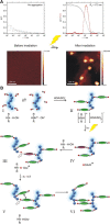
A light-switchable electrostatically self-assembled system with photo addressable particle size; top: schematic representation. Starting from a divalent anionic azobenzene dye and cationic dendrimeric macroions, self-assembled nanoparticles result (A), which grow in size upon irradiation with UV light (B); middle: AFM (A) before and (B) after UV irradiation and (C) corresponding DLS for a loading ratio l = 4.5; (D) Isothermal titration calorimetry revealing the different binding enthalpies of the cis and the trans dye isomer; (E) ζ-potential for dendrimer–dye assemblies before and after irradiation for different loading ratios confirming the charge density control of the particle size (Willerich and Gröhn, 2010; Willerich and Gröhn, 2011b). Reprinted with permission. Copyright © 2011 American Chemical Society and © 2010 WILEY-VCH Verlag GmbH and Co. KGaA, Weinheim.
FIGURE 20
FIGURE 20
{Mo72V30}–azo complex with irradiation dependent interconnection; top: schematic representation; bottom: SEM of dry films prepared by drop-casting (A) {Mo72V30} before functionalization with azo molecules; (B) the {Mo72V30}-trans-azo hybrid structure; (C) {Mo72V30}-cis-azo obtained upon UV-light irradiation (365 nm) prior to drop-casting of the {Mo72V30}-trans-azo solution (Markiewicz et al., 2017). Reprinted with permission. Copyright © The Royal Society of Chemistry 2017.
FIGURE 21
FIGURE 21
Light switchable nano-object shape by electrostatic self-assembly: An anionic azo dye (Ay38) and a linear flexible polyelectrolyte (PDADMA) for micrometer long thin fibers which compact into ellipsoids upon irradiation (Mariani et al., 2018). Reprinted with permission. Copyright © 2018 WILEY-VCH Verlag GmbH and Co. KGaA, Weinheim.
FIGURE 22
FIGURE 22
Study of the formation mechanism in light-responsive electrostatic self-assembly: (A) Selected SANS profiles of reforming PA–diAzoEt complexes after irradiation and reassembly at 45°C. The solid lines represent fits to the model of a mixture of free polymer chains and complexes; (B) Illustration of the mechanism of the self-assembly of the PA−diAzoEt complexes (Carl et al., 2020). Reprinted with permission. Copyright © 2019 American Chemical Society.
FIGURE 23
FIGURE 23
Light-triggerable enzyme aggregation and activity; top: Schematic representation of the assemblies of an ionic spiropyrane and lysozyme; bottom: Interconversion of the two spiropyran isomers in inverse photochromism (Moldenhauer et al., 2019). Reprinted with permission. Copyright © 2018 American Chemical Society.
FIGURE 24
FIGURE 24
Photoacid which changes the pKa in the excited state acting as a building block in electrostatic self-assembly with linear oligo (ethylene imine): A DLS and AFM before (left) and after (right) UV irradiation. B: light-induced assembly mechanism (Cardenas-Daw and Gröhn, 2015). Reprinted with permission. Copyright © 2015 American Chemical Society.
FIGURE 25
FIGURE 25
Formation of ETAB/Eu-POM supramolecular materials and their responsiveness to UV Light, Cu2+, and H+. (Guo et al., 2016b). Reprinted with permission. Copyright © 2016 American Chemical Society.
FIGURE 26
FIGURE 26
A ternary multiswitchable assembly formed of Flavy, a photoacid (1N36S) and linear poly (allylamine) (simplified schematic representation displaying one molecule of each component per assembly only). (A): The protonated photoacid and Flavy in its closed form. (B): The deprotonated photoacid and hydroxylated Flavy. (C): The protonated photoacid and protonated Flavy. (D): The protonated photoacid and hydroxylated Flavy. (E): The deprotonated photoacid and open Flavy. (F): The deprotonated photoacid and hydroxylated Flavy (Zika and Gröhn, 2021). The Figure was reproduced, © 2021 A. Zika and F. Gröhn, distributed under the terms of the Creative Commons Attribution 4.0 International License.
FIGURE 27
FIGURE 27
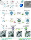
Polyelectrolyte brush-porphyrin assemblies as photocatalysts: (A) Scheme, and UV/Vis absorption spectra in dependence on irradiation time for catalyzing the iodide oxidation as the model reaction: (B) TMPyP without polyelectrolyte and (C) a TMPyP/PSS brush sample; The bands at λ = 353 nm and at λ = 287 nm indicate the faster tri-iodide formation with the nano-assembly as the catalyst (Frühbeißer and Gröhn, 2012). Reprinted with permission. Copyright © 2012 American Chemical Society.
FIGURE 28
FIGURE 28
TAPP-G4 dendrimer aggregates and the dependence of the catalytic activity on the internal stacking of the porphyrins: (A) UV/Vis analysis in dependence on the loading ratio l: shift of the Soret band and changes in the extinction coefficient ε of the Soret band at pH 11; inset: Soret band; (B) fluorescence analysis in dependence on the loading ratio l: area of the fluorescence peak at 574 nm ≤ λ ≤ 800 nm; inset: fluorescence; (C) Schematic illustration of TAPP−G7.5 dendrimer assemblies with l = 0.05, l = 1.0, and l = 1.6 (red: TAPP molecules; blue: G7.5 dendrimers); (D) photocatalytic degradation of methyl orange upon irradiation with visible light similar to the sun spectrum as a photocatalytic model reaction: Decrease of the methyl orange concentration measured at λ = 464 nm for TAPP−G7.5 dendrimer assemblies with different loading ratios and for TAPP only (Krieger et al., 2017). Reprinted with permission. Copyright © 2017 American Chemical Society.
FIGURE 29
FIGURE 29
Photodegradation of Rose Bengal by porphyrin-porphyrin microrhombuses decorated with ZnO nanorods: (A) Time-dependent UV/Vis spectroscopy of the degradation of Rose Bengal with the ZnO microrhombuses. (B) Catalytic degradation of Rose Bengal with ZnO-microrhombuses (blue circles), with TMPyP (red rhombs), with TPPS (green squares), with ZnO-nanorods (gray inverted triangles), and without catalyst (black triangles) (Düring et al., 2017b). Reprinted with permission. Copyright © 2017 WILEY-VCH Verlag GmbH and Co. KGaA, Weinheim.
FIGURE 30
FIGURE 30
POM–dendrimer self-assembly and photocatalysis in aqueous solution as investigated: (A) Scheme and photos of the photocatalytic degradation of methyl red by POM–dendrimer assemblies and POM in aqueous solution; A: POM–dendrimer before irradiation, (B): POM–dendrimer after 20 min UV-irradiation, (C): POM before irradiation, (D): POM after 20 min UV-irradiation; The dye degrades much more with the POM–dendrimer assembly (A → B) as compared to the cluster only solution (C → D) (Kutz et al., 2018). Reprinted with permission. Copyright © The Royal Society of Chemistry 2018.

Similar articles

Cited by

References

    1. Achilleos D. S., Hatton T. A., Vamvakaki M. (2012). Light-Regulated Supramolecular Engineering of Polymeric Nanocapsules. J. Am. Chem. Soc. 134, 5726–5729. 10.1021/ja212177q - DOI - PubMed
    1. Agmon N., Rettig W., Groth C. (2002). Electronic Determinants of Photoacidity in Cyanonaphthols. J. Am. Chem. Soc. 124, 1089–1096. 10.1021/ja003875m - DOI - PubMed
    1. Amaral S. P., Tawara M. H., Fernandez-Villamarin M., Borrajo E., Martínez-Costas J., Vidal A., et al. (2018). Tuning the Size of Nanoassembles: A Hierarchical Transfer of Information from Dendrimers to Polyion Complexes. Angew. Chem. Int. Ed. 57, 5273–5277. 10.1002/anie.201712244 - DOI - PubMed
    1. Andrade S. M., Costa S. M. B. (2002). Spectroscopic Studies on the Interaction of a Water Soluble Porphyrin and Two Drug Carrier Proteins. Biophysical J. 82, 1607–1619. 10.1016/s0006-3495(02)75512-4 - DOI - PMC - PubMed
    1. Anraku Y., Kishimura A., Oba M., Yamasaki Y., Kataoka K. (2010). Spontaneous Formation of Nanosized Unilamellar Polyion Complex Vesicles with Tunable Size and Properties. J. Am. Chem. Soc. 132, 1631–1636. 10.1021/ja908350e - DOI - PubMed