Review
. 2022 Jan 27;3(3):269-287.
doi: 10.1039/d1cb00195g. eCollection 2022 Mar 9.

Chemogenetics of cell surface receptors: beyond genetic and pharmacological approaches

Affiliations
Review

Chemogenetics of cell surface receptors: beyond genetic and pharmacological approaches

Yuta Miura et al. RSC Chem Biol. .

Abstract

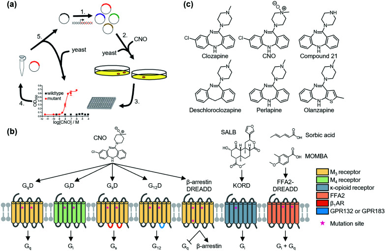
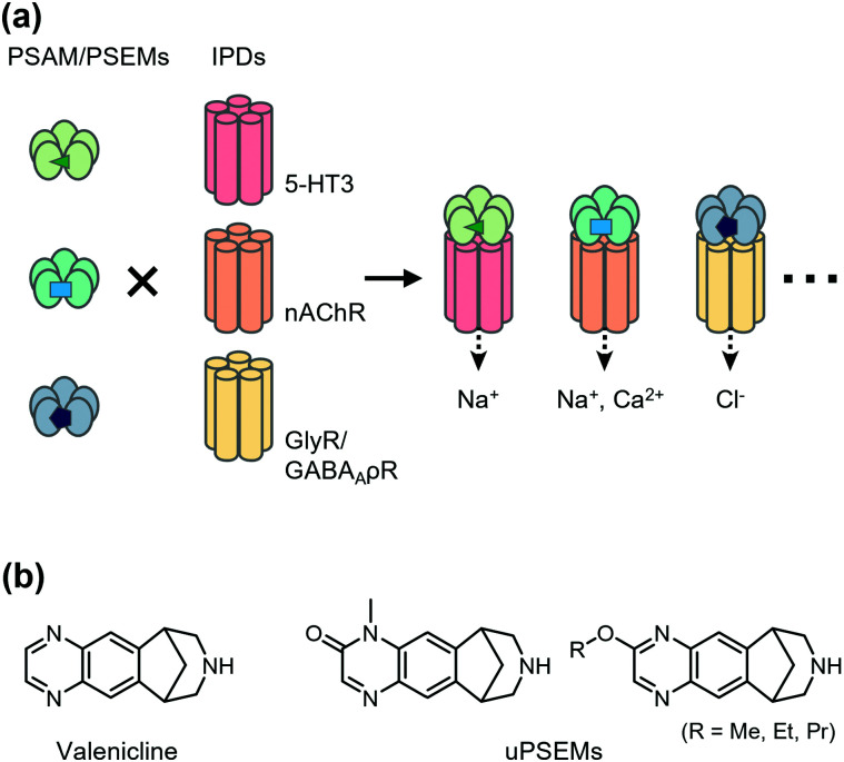
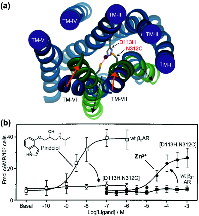
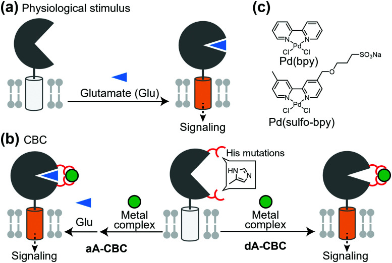
Cell surface receptors transmit extracellular information into cells. Spatiotemporal regulation of receptor signaling is crucial for cellular functions, and dysregulation of signaling causes various diseases. Thus, it is highly desired to control receptor functions with high spatial and/or temporal resolution. Conventionally, genetic engineering or chemical ligands have been used to control receptor functions in cells. As the alternative, chemogenetics has been proposed, in which target proteins are genetically engineered to interact with a designed chemical partner with high selectivity. The engineered receptor dissects the function of one receptor member among a highly homologous receptor family in a cell-specific manner. Notably, some chemogenetic strategies have been used to reveal the receptor signaling of target cells in living animals. In this review, we summarize the developing chemogenetic methods of transmembrane receptors for cell-specific regulation of receptor signaling. We also discuss the prospects of chemogenetics for clinical applications.


Conflict of interest statement

There are no conflicts to declare.

Figures

Fig. 1
Fig. 1. Schematic illustration of DREADDs. (a) Development of DREADD by directed molecular evolution. Reproduced with permission from ref. . Copyright 2007 by Proceedings of the National Academy of Sciences. (b) Structure and G protein-coupling properties of DREADDs. All DREADDs are unable to bind to the endogenous agonists, but can be activated by synthetic ligands. (c) Chemical structures of clozapine, CNO, and new muscarinic receptor-based DREADD ligands.
Fig. 2
Fig. 2. Schematic illustration of PSAM/PSEM system. (a) By combining different IPDs for a given PSAM/PSEM pair, multiple functional outcomes can be achieved, including neuronal activation, regulation of calcium flux, and neuronal silencing. Development of multiple orthogonal PSAM/PSEM pairs has enabled the combinatorial generation of diverse chemogenic ion channel tools. (b) Chemical structures of varenicline and uPSEMs.
Fig. 3
Fig. 3. Metal-ion-induced activation of a β2AR mutant. (a) The active form of the receptor is stabilized by the binding of metal ions to the introduced coordination sites (D113H/N312C). (b) β2AR(D113H/N312C) is not activated by normal agonists, but is activated by metal ions. Reproduced with permission from ref. and . Copyright 2006 by the American Society for Biochemistry and Molecular Biology and 1999 by Proceedings of the National Academy of Sciences.
Fig. 4
Fig. 4. Schematic illustration of coordination-based chemogenetics. (a) Glutamate-binding induces the closing of the LBD for activation of the receptors. (b) Coordination-based chemogenetics (CBC) for glutamate receptors. In aA-CBC (allosteric activation via coordination-based chemogenetics), metal complexes act as PAM. In dA-CBC, metal complexes act as direct activator without affecting sensitivity to glutamate. (c) Chemical structures of Pd(bpy) and its derivative, Pd(sulfo-bpy) showing low cytotoxicity.
Fig. 5
Fig. 5. Ligand-induced dimerization and its applications to membrane receptors. (a) Chemical structure of rapamycin (Rap). Black or red region correspond to those interacting with FKBP or FRB, respectively. (b) Schematic illustration of intracellular heterodimerization of TGF-βRI and TGF-βRII by rapamycin. (c) Application of heterodimerization into AND logic gate for the precise control of CAR-T cell. In this system, both antigen-recognition and addition of the dimerizer are required for the signal transduction. (d) Ligand-induced translocation of Gα protein to transmembrane for dissecting its role in the heterotrimeric complex. ER, endoplasmic reticulum.
Fig. 6
Fig. 6. Application of light-induced dimerization or oligomerization to membrane receptors. (a) Representative systems of light-inducible dimerization or oligomerization. (b) Expansion of wavelength available for photoactivate proteins. Three black bars correspond the wavelength covered by each chromophore. (c) Regulation of RTK with light. Intracellular region of RTK is fused with PHR domain of CRY2, which homodimerizes upon photo-irradiation. TKD, Tyrosine kinase domain. (d) Photo-Induced interaction between β2AR and β-arrestin utilizing CRY2-CIB interaction.
Fig. 7
Fig. 7. Photoswitching of PTL-tethered receptors. (a) Chemical structures of various azobenzene-based photoswitchable motifs. (b) Chemical structures and the photoisomerization of diazocines and arylazopyrazoles. (c) Chemical structure of a PTL, MAQ. (d) Chemical structure of MAG and schematic illustration of a light-responsive ionotropic glutamate receptor, LiGluK2. (e) Restoration of pupillary reflex in TKO mice lacking phototransduction and pupillary reflexes by expression LiGluK2 (right). Representative infrared images of the pupil area taken in the dark (left column) and under 380 nm light (right column) for WT, TKO, and TKO-LiGluK2 mice are shown. Reproduced with permission from ref. . Copyright 2011 by Elsevier, Inc.
Fig. 8
Fig. 8. Optical control of a PORTL (BGAG12)-tethered mGlu2 (SNAG-mGlu2). (a) Schematic illustration of PORTL-mediated reversible regulation of SNAG-mGlu2 (top). Chemical structure of the PORTL, BGAG12 (bottom). (b) Schematic illustration of branched BGAG12-tethered mGlu2 (left). Bar plot showing photoswitch efficiencies for different levels of branched BGAG12-tethered mGlu2 (right). Reproduced with permission from ref. . Copyright 2020 by Elsevier, Inc. (c) Chemical structure of an optimized PORTL with 4-urea-4′-amide azobenzene, BGAG12,400.
Fig. 9
Fig. 9. Non-covalent ligand tethering for chemogenetic regulation of receptors of interest. (a) Schematic illustration of non-covalent tethering of photoswitchable ligands using anti-GFP NPCs. NB, nanobody. (b) Schematic illustration of non-covalent agonist tethering using metal coordination for the selective activation of GPCRs of interest.
Fig. 10
Fig. 10. General principle of genetic code expansion. (a) Schematic illustration of the incorporation of UAAs into transmembrane receptors. RS, orthogonal aminoacyl-tRNA synthetase. (b–e) Chemical structures of representative UAA structures for photo-crosslinking (in b), photo-cleavage (in c), photo-switching (in d) and bioorthogonal tethering (in e).
Fig. 11
Fig. 11. Photoswitching of NMDARs using genetic-code expansion. (a) Structure of NMDARs composed of GluN1 and GluN2 subunits. In the right panel, the top view of the LBD is shown, and mutation site (P523) is highlighted. (b) Structure of the transmembrane domain (TMD) of NMDARs represented by a side (Left) and top view (Right). (c) Representative current traces of NMDARs having an azobenzene-based photo-responsive UAA (PSAA) at P532 in the LBD (in left) or at F654 in the TMD (in right). Reproduced with permission from ref. . Copyright 2017 by eLife Sciences Publications, Ltd.
Fig. 12
Fig. 12. Schematic illustration of t-toxin and lumitoxin. (a) T-Toxin inhibits the target membrane receptor in a cell-specific manner. (b) Lumitoxin having LOV2 domain can reversibly control the target receptor by photo-irradiation.
Fig. 13
Fig. 13. DART and maPORTL system for regulation of endogenous receptors. (a) Chemical structure of a designer ligand for DART system for AMPAR. (b) Schematic illustration of DART system for cell-specific inhibition of endogenous AMPARs. (c) Chemical structure of maPORTL ligand for mGlu2. (d) Schematic illustration of maPORTL system for photoswitching of endogenous mGlu2.
Fig. 14
Fig. 14. Chemogenetic regulation of endogenous NMDAR using prodrug approach. (a) Chemical structure of a PLE-specific prodrug for NMDAR. (b) Schematic illustration of cell-specific inhibition of NMDAR using prodrug. The prodrug is converted into active inhibitor, MK801 to inhibit NMDAR in a cell-specific manner by PLE.
None
Yuta Miura
None
Akinobu Senoo
None
Tomohiro Doura
None
Shigeki Kiyonaka

References

    1. Anzalone A. V. Koblan L. W. Liu D. R. Nat. Biotechnol. 2020;38:824–844. doi: 10.1038/s41587-020-0561-9. - DOI - PubMed
    1. Gierut J. J. Jacks T. E. Haigis K. M. Cold Spring Harb. Protoc. 2014;2014:339–349. - PMC - PubMed
    1. Fenno L. Yizhar O. Deisseroth K. Annu. Rev. Neurosci. 2011;34:389–412. doi: 10.1146/annurev-neuro-061010-113817. - DOI - PMC - PubMed
    1. Boyden E. S. Zhang F. Bamberg E. Nagel G. Deisseroth K. Nat. Neurosci. 2005;8:1263–1268. doi: 10.1038/nn1525. - DOI - PubMed
    1. Airan R. D. Thompson K. R. Fenno L. E. Bernstein H. Deisseroth K. Nature. 2009;458:1025–1029. doi: 10.1038/nature07926. - DOI - PubMed