. 2022 Mar 14:9:835976.
doi: 10.3389/fmolb.2022.835976. eCollection 2022.

Degenerative Nucleus Pulposus Cells Derived Exosomes Promoted Cartilage Endplate Cells Apoptosis and Aggravated Intervertebral Disc Degeneration

Affiliations

Degenerative Nucleus Pulposus Cells Derived Exosomes Promoted Cartilage Endplate Cells Apoptosis and Aggravated Intervertebral Disc Degeneration

Xiaofei Feng et al. Front Mol Biosci. .

Abstract

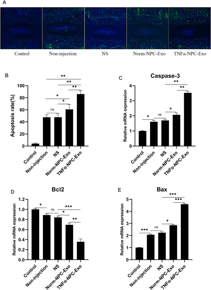
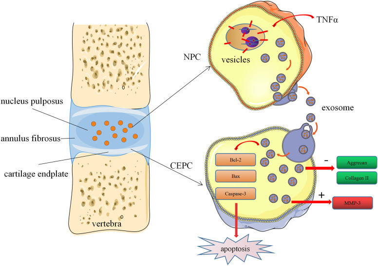
Intervertebral disc (IVD) degeneration is a complex multifactorial disease model, which pathogenesis has not been fully defined. There are few studies on the information interaction between nucleus pulposus (NP) cells and cartilage endplate (CEP) cells. Exosomes, as a carrier of information communication between cells, have become a research hotspot recently. The purpose of this study was to explore whether degenerative NP cells-derived exosomes promoted CEP cells apoptosis and aggravated IVD degeneration. The degenerative NP cells model was induced by TNFα. NPC exosomes were isolated from the supernatant of the NP cell culture medium. The viability of NP cells and CEP cells was examined by CCK-8 assays. The exosomes were identified by TEM, NTA, and western blot. Extracellular matrix (ECM) metabolism was measured by cellular immunofluorescence and qRT-PCR. Apoptosis was detected by flow cytometry and TUNEL. X-ray and magnetic resonance imaging (MRI), as well as hematoxylin-eosin (H&E), Safranine O-Green staining was adopted to evaluate IVD degeneration grades. TNFα had a minor impact on NPC viability but inhibited ECM synthesis and promoted ECM degradation. TNFα-NPC-Exo had less effect on CEPC proliferation but promoted CEPC apoptosis and affect ECM metabolism, inhibiting aggrecan and collagen II expression and enhancing MMP-3 expression. TNFα-NPC-Exo aggravates IVD degeneration in a rat model and promoted CEPC apoptosis. In conclusion, this study demonstrated that degenerated NPC-exosome could induce apoptosis of CEPCs, inhibit ECM synthesis, and promote ECM degradation. In addition, it was proved that degenerated NPC-exosome aggravates IVD degeneration.

Keywords: apoptosis; cartilage endplate; exosome; intervertebral disc degeneration; nucleus pulposus.


Conflict of interest statement

The authors declare that the research was conducted in the absence of any commercial or financial relationships that could be construed as a potential conflict of interest.

Figures

FIGURE 1
FIGURE 1
Effect of TNFα on the viability and ECM metabolism of NPCs. (A) NPCs were treated with 0, 1, 5, 10, 20, 30, 50 ng/ml TNFα for 24 h and detected by CCK-8 assay (n = 3). (B) Typical images of immunofluorescence of Aggrecan in NPCs photographed by fluorescence microscopy (scale bar = 100 μm). (C) Typical images of immunofluorescence of Collagen II in NPCs photographed by fluorescence microscopy (scale bar = 100 μm). (D) Typical images of immunofluorescence of MMP3 in NPCs photographed by fluorescence microscopy (scale bar = 100 μm). (E) Semi-quantitative analysis of Aggrecan, Collagen II, and MMP-3 fluorescence intensity (cells = 100). (F) The expression levels of Aggrecan, Collagen II, and MMP-3 were analyzed by qRT-PCR in NPCs, which were cocultured for 24 h with 5 ng/ml TNFα. All results are representative of at least three independent experiments and each value is the mean ± s.d. of three determinations. *p < 0.05, **p < 0.01 and ***p < 0.001.
FIGURE 2
FIGURE 2
Identification and characterization of nucleus pulposus-derived exosomes and the uptake of exosomes in CEPCs. (A) Transmission electron micrograph of purified particles. The image showed small vesicles of approximately 100 nm in diameter (scale bar = 100 nm). (B) Size distribution of vesicles secreted by NPCs determined by NTA. The average particle size was 153.6 nm. (C) Expression of exosomes markers (CD9, CD63, and TSG101) and nuclear marker (Calnexin) detected by Western blot. The protein expression of exosomes markers was detectable in NPC exosomes but not NPCs. (D) NPCs were cultured under normal medium and 5 ng/ml TNFα conditions for 24 h, and the protein expression of exosomes was measured by immunoblot. TNFα increased the production of exosomes in NPCs. (E) The uptake of NPC exosome in CEPC. Compared with co-culture for 6 h, a large number of NPC exosomes were taken up in CEPCs after co-culture for 24 h. All results are representative of at least three independent experiments and each value is the mean ± s.d. of three determinations. *p < 0.05, **p < 0.01 and ***p < 0.001.
FIGURE 3
FIGURE 3
Effects of Norm-NPC-Exo and TNFα-NPC-Exo on proliferation and apoptosis of CEPCs. (A) CEPCs were treated with 0, 10, 20, 30, 40, 50 μg/ml Norm-NPC-Exo and TNFα-NPC-Exo for 24 h and detected by CCK-8 assay (n = 3). (B) CEPCs were treated with 20nμg/ml Norm-NPC-Exo and TNFα-NPC-Exo for 0, 2, 4, 6, 8, 10, 12 days and detected by CCK-8 assay (n = 3). (C) The cell viability of different groups of CEPC was calculated according to OD value. The results showed that there was no significant difference in the cell proliferation multiple among the three groups. (D) The percentage of apoptotic CEPCs could be increased to 3.88 ± 0.05% at 24 h or to 5.16 ± 0.04% at 48 h on TNFα-NPC-Exo treated CEPCs. Compared with the control and Norm-NPC-Exo group, TNFα-NPC-Exo causes a significant change in apoptosis rate. (E) CEPCs were treated with Normal medium, Norm-NPC-Exo, and TNFα-NPC-Exo for 24 and 48 h. Representative dot plots of apoptosis flow cytometry detection were shown. (F) Western blot analyzed caspase-3, cleaved caspase-3, Bax, and Bcl-2 in CEPCs after treatment of different exosomes. (G) Semi-quantitative analysis of caspase-3, cleaved caspase-3, Bax, and Bcl-2 levels (n = 3). All results are representative of at least three independent experiments and each value is the mean ± s.d. of three determinations. *p < 0.05, **p < 0.01.
FIGURE 4
FIGURE 4
Effects of Norm-NPC-Exo and TNFα-NPC-Exo on ECM metabolism of CEPCs. (A) CEPCs were treated with 20 μg/ml Norm-NPC-Exo and TNFα-NPC-Exo for 24 h. Representative images of immunofluorescence of aggrecan, collagen II and MMP-3 in CEPCs photographed by fluorescence microscopy (scale bar = 100 μm). (B) Semi-quantitative analysis of aggrecan, collagen II, and MMP-3 fluorescence intensity (n = 3). Compared with the control and Norm-NPC-Exo group, the expression of aggrecan and collagen II in CEPC cytoplasm was significantly decreased at TNFα-NPC-Exo group, while the expression of MMP-3 was significantly increased. (C) The expression levels of aggrecan, collagen II and MMP-3 were analyzed by qRT-PCR in CEPCs. Aggrecan and collagen II expression were significantly decreased following TNFα-NPC-Exo treatment, while MMP-3 expression was significantly increased. All results are representative of at least three independent experiments and each value is the mean ± s.d. of three determinations. *p < 0.05, **p < 0.01 and ***p < 0.001.
FIGURE 5
FIGURE 5
Intradiscal injection of TNFα-NPC-Exo alleviated the IVD degeneration in a rat model. (A) Modeling method of rat caudal vertebra by acupuncture. (B) Representative images of X-ray film were obtained at 2, 4, and 6 weeks after needle puncture. (C) Representative images of MRI film were obtained at 2, 4, and 6 weeks after acupuncture. (D) Changes in disc height index (DHI) of the indicated groups after a puncture. The DHI was measured at weeks 2, 4, 6 timing. A significant decrease of the DHI was observed in all puncture groups at 2 weeks after surgery. And the more serious decrease of the DHI was noted in TNFα-NPC-Exo groups (n = 8). (E) The change of MRI grade in the indicated groups after a puncture. The degree of disc degeneration by MRI grade was significantly higher in the TNFα-NPC-Exo groups than in the non-injection group (n = 8). (F) Representative images of HE staining. (G) Representative images of Safranine O-Green staining. Each value are expressed as mean ± s.d *p < 0.05, **p < 0.01, and ***p < 0.001.
FIGURE 6
FIGURE 6
Intradiscal injection of TNFα-NPC-Exo promoted the CEPC apoptosis in a rat model. (A) TUNEL staining of IVDs in the indicated groups at 6 weeks after needle puncture. Green fluorescence (FITC) indicating TUNEL-positive cells. Blue fluorescence (DAPI) indicates total cells (scale bar = 150 μm). (B) A significant increase in the apoptosis rate was observed in the TNFα-NPC-Exo group compared with the non-injection group. (C) qRT-PCR showed that there are significantly increased levels of caspase-3 in the punctured IVDs by the injection of TNFα-NPC-Exo compared with other groups. (D) The mRNA expression of Bcl-2 in CEPCs was detected using qRT-PCR. Bcl-2 expression was significantly decreased following TNFα-NPC-Exo treatment. (E) The mRNA expression of Bax in CEPCs was detected using qRT-PCR. Bax expression was significantly increased following TNFα-NPC-Exo treatment. All results are representative of at least three independent experiments and each value is the mean ± s.d. of three determinations. *p < 0.05, **p < 0.01 and ***p < 0.001.
FIGURE 7
FIGURE 7
Schematic of our working hypothesis. Degenerated NPC-derived exosomes induce apoptosis of CEPCs, inhibit ECM synthesis, and promote ECM degradation.

Similar articles

Cited by

References

    1. Adams M. A., Roughley P. J. (2006). What Is Intervertebral Disc Degeneration, and what Causes it? Spine (Phila Pa 1976) 31 (18), 21512161. 10.1097/01.brs.000023176 - DOI - PubMed
    1. Ailawadi S., Wang X., Gu H., Fan G.-C. (2015). Pathologic Function and Therapeutic Potential of Exosomes in Cardiovascular Disease. Biochim. Biophys. Acta (Bba) - Mol. Basis Dis. 1852 (1), 1–11. 10.1016/j.bbadis.2014.10.008 - DOI - PMC - PubMed
    1. Cazzanelli P., Wuertz-Kozak K. (2020). MicroRNAs in Intervertebral Disc Degeneration, Apoptosis, Inflammation, and Mechanobiology. Int. J. Mol. Sci. 21 (10), 3601. 10.3390/ijms21103601 - DOI - PMC - PubMed
    1. Che Y. J., Guo J. B., Liang T., Chen X., Zhang W., Yang H. L., et al. (2019). Assessment of Changes in the Micro-nano Environment of Intervertebral Disc Degeneration Based on Pfirrmann Grade. Spine J. 19, 1242–1253. 10.1016/j.spinee.2019.01.008 - DOI - PubMed
    1. Chen S., Fu P., Wu H., Pei M. (2017). Meniscus, Articular Cartilage and Nucleus Pulposus: a Comparative Review of Cartilage-like Tissues in Anatomy, Development and Function. Cell Tissue Res 370 (1), 53–70. 10.1007/s00441-017-2613-0 - DOI - PMC - PubMed