. 2022 Mar 11:13:832928.
doi: 10.3389/fphar.2022.832928. eCollection 2022.

Hypericum lanceolatum Lam. Medicinal Plant: Potential Toxicity and Therapeutic Effects Based on a Zebrafish Model

Affiliations

Hypericum lanceolatum Lam. Medicinal Plant: Potential Toxicity and Therapeutic Effects Based on a Zebrafish Model

Laura Gence et al. Front Pharmacol. .

Abstract

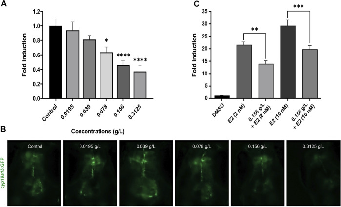
Hypericum lanceolatum Lam. (H. lanceolatum) is a traditional medicinal plant from Reunion Island used for its pleiotropic effects mainly related to its antioxidant activity. The present work aimed to 1) determine the potential toxicity of the plant aqueous extract in vivo and 2) investigate its putative biological properties using several zebrafish models of oxidative stress, regeneration, estrogenicity, neurogenesis and metabolic disorders. First, we characterized the polyphenolic composition by liquid chromatography-tandem mass spectrometry (LC-MS/MS) and identified chlorogenic acid isomers, quercetin and kaempferol derivatives as the major compounds. We then evaluated for the first time the toxicity of an aqueous extract of H. lanceolatum and determined a maximum non-toxic concentration (MNTC) in zebrafish eleutheroembryos from 0 to 96 hpf following OECD (Organization for Economic Cooperation and Development) guidelines. This MNTC test was also determined on hatched eleutheroembryos after 2 days of treatment (from 3 to 5 dpf). In our study, the anti-estrogenic effects of H. lanceolatum are supported by the data from the EASZY assay. In a tail amputation model, we showed that H. lanceolatum at its MNTC displays antioxidant properties, favors immune cell recruitment and tissue regeneration. Our results also highlighted its beneficial effects in metabolic disorders. Indeed, H. lanceolatum efficiently reduces lipid accumulation and body mass index in overfed larva- and adult-models, respectively. In addition, we show that H. lanceolatum did not improve fasting blood glucose levels in a hyperglycemic zebrafish model but surprisingly inhibited neurogenesis impairment observed in diabetic conditions. In conclusion, our study highlights the antioxidant, pro-regenerative, anti-lipid accumulation and pro-neurogenic effects of H. lanceolatum in vivo and supports the use of this traditional medicinal plant as a potential alternative in the prevention and/or treatment of metabolic disorders.

Keywords: LC MS/MS; Toxicity; Zebrafish; diabetes; neurogenesis; obesity; regeneration.


Conflict of interest statement

The authors declare that the research was conducted in the absence of any commercial or financial relationships that could be construed as a potential conflict of interest.

Figures

FIGURE 1
FIGURE 1
Spectra obtained after characterization of H. lanceolatum aqueous extract by LC-MS/MS. (A) Representative total ion chromatogram (TIC) obtained in negative mode (B) HPLC-UV chromatogram obtained at 280 nm.
FIGURE 2
FIGURE 2
In vivo acute toxicity assays in zebrafish embryos and eleutheroembryos. Toxicity curves, estimated median lethal concentrations (LC50) and representative pictures of (A) 96-hpf embryos and (B) 5-dpf eleutheroembryos exposed to different concentrations of H. lanceolatum aqueous extract or E3 medium (Control). Curve values are expressed as means ± SEM (n = 60 animals/group from 3 independent experiments). ***p < 0.001 (Student t-test). Bar = 350 µm. Note that in (A), at 5 g/L all the embryos are dead at 24-hpf. For 2.5 and 1.25 g/L around 26 and 63% of eleutheroembryos were alive at 96-hpf, respectively. The shaded area represents the MNTC.
FIGURE 3
FIGURE 3
H. lanceolatum did not impact the expression of toxicity and endocrine disruption biomarkers. (A) qPCR analysis of recognized biomarkers for hepatotoxicity (fabp10a, gclc), cardiotoxicity (erg), nephrotoxicity (ctgf) and glucocorticoid signaling (fkpb5) of eleutheroembryos treated with H. lanceolatum aqueous extract at the MNTC (0.3125 g/L) or E3 medium (Control) for 2 days (n = 7–8 pools of 20 eleutheroembryos/group). (B) Fluorescence quantification of spinal cord in tg (GFAP::GFP) eleutheroembryos (dotted rectangle) and representative pictures of 48 h-treated eleutheroembryos (n = 35/group from 3 independent experiments). Results are expressed as means ± SEM. Bar = 200 µm.
FIGURE 4
FIGURE 4
H. lanceolatum aqueous extract did not impact the behavioral activity in larvae. (A) Graphs showed the inactivity, small activity, large activity and total distances of 6-dpf larvae after 3 days of incubation with the H. lanceolatum aqueous extract at the MNTC (HL) or E3 medium (Control) (B) Total distance of control and treated larvae. Individual values were represented with means ± SEM (n = 11–12 animals/group). Bar = 2.3 mm.
FIGURE 5
FIGURE 5
H. lanceolatum aqueous extract possesses an anti-estrogenic activity. (A) Quantification of GFP fluorescence and (B) representative pictures of embryos exposed to water (Control) and different concentrations of H. lanceolatum aqueous extract (g/L) for 96 h (n = 26–38/group from 2 independent experiments). * denotes significant differences as compared to the control group (Dunn’s multiple comparison test, *p = 0.0438 and ****p < 0.0001). (C) Fluorescence quantification of GFP in embryos treated with control solvent (DMSO) and E2 at two different concentrations (2 and 10 nM) alone or co-exposed with H. lanceolatum aqueous extract 0.156 g/L (n = 19–21/group). * denotes significant differences between groups (Dunnett’s multiple comparison test, **p = 0.0011 and ***p = 0.0002). All the results are expressed as fold induction above control (means ± SEM).
FIGURE 6
FIGURE 6
H. lanceolatum aqueous extract exhibits preventive antioxidant effect after caudal fin amputation. Fluorescence quantification and representative pictures of oxidized (A) DCFH-DA and (B) DHE probes after caudal fin amputation of adult zebrafish pre-incubated in water (Control) or H. lanceolatum aqueous extract at the MNTC (0.3125 g/L H. lanceolatum). Results are expressed as means ± SEM (n = 9/group from 3 independent experiments). **p < 0.01 (Student t-test). Bar = 200 µm.
FIGURE 7
FIGURE 7
H. lanceolatum aqueous extract improves the recruitment of neutrophils and macrophages after tail injury. Number of (A) Lcp1-positive (macrophage) and (B) Mpo-positive (neutrophils) cells (arrows) at the injury site (white box) and representative pictures of tail amputations of 3-dpf eleutheroembryos, incubated with E3 medium (Control) or H. lanceolatum aqueous extract at 0.3125 g/L (H. lanceolatum) for 6 h. Dotted lines represent the tail amputation level. Results are expressed as means ± SEM (n = 34/group from 3 independent experiments). **p < 0.01, ***p < 0.001 (Student t-test). Bar = 40 µm.
FIGURE 8
FIGURE 8
H. lanceolatum aqueous extract improves tail regeneration in zebrafish eleutheroembryos. (A) Measurement of tail regeneration area (in arbitrary units) and representative pictures of eleutheroembryos incubated in E3 medium (Control) or H. lanceolatum aqueous extract at 0.3125 g/L (H. lanceolatum), 0- and 3-days post-amputation (dotted) of 3-dpf eleutheroembryos. (B) Measurement of tail regeneration area and representative pictures of eleutheroembryos treated with the vehicle (0.4% ethanol), dexamethasone (500µM, DEX) or H. lanceolatum aqueous extract + dexamethasone (H. lanceolatum + DEX), 0- and 3-days post-amputation. Results are expressed as means ± SEM (n = 20–39/group from 3 independent experiments). **p < 0.01, ***p < 0.001 (Student t-test). Bar = 25 µm.
FIGURE 9
FIGURE 9
H. lanceolatum aqueous extract displays pro-regenerative properties in the tail of adult fish. (A) Measurement of tail regeneration area of adult fish after a 14-days incubation period with fish water (Control) or H. lanceolatum aqueous extract at 0.3125 g/L (H. lanceolatum). (B) Representative pictures of caudal fin regeneration 3-, 7-, and 14-days post-amputation. Dotted lines represent the tail amputation level. Results are expressed as means ± SEM (n = 4–7 animals/group). *p < 0.05, ***p < 0.001 (Student t-test). Bar = 625 µm.
FIGURE 10
FIGURE 10
H. lanceolatum aqueous extract inhibits lipid accumulation in a HFD model of larvae. (A) Quantification of ORO staining and (B) representative pictures of normally fed larvae (Control), overfed (HFD) and overfed larvae treated with H. lanceolatum aqueous extract at 0.3125 g/L (HFD + HL). Dotted lines indicate the ORO staining in the digestive tract. Arrows show the staining in vessels including dorsal aorta (1), posterior cardinal vein (2) and caudal vein (3). Results are expressed as means ± SEM (n = 45 larvae/group from 3 independent experiments). ***p < 0.001 (One-way ANOVA). Bar = 150 µm.
FIGURE 11
FIGURE 11
H. lanceolatum aqueous extract induces weight loss properties in DIO model of adult fish. (A) Body mass index (BMI) at day-14 and (B) body weight of normally fed (Control), overfed (DIO) and overfed fish treated with H. lanceolatum (DIO + HL). Results are expressed as means ± SEM (n = 8–10/group). *p < 0.05, **p < 0.01 (One-way ANOVA).
FIGURE 12
FIGURE 12
H. lanceolatum aqueous extract has “anti-diabetic” effects by maintaining homeostatic neurogenesis in chronic hyperglycemic adult fish. (A) Fasting blood glucose levels of control (Control), chronic hyperglycemic (CHG) and chronic hyperglycemic fish treated with H. lanceolatum (CHG + HL). (B) Number of PCNA-positive cells according to the studied neurogenic regions. (C) Representative pictures of proliferative activity (PCNA) in zebrafish brains. (D) Total number of PCNA-positive cells, counted in all the studied regions, in Control, CHG and CHG + HL-treated fish. Dm, medial zone of dorsal telencephalic area. PPa, parvocellular preoptic nucleus, anterior part. Hv, ventral zone of periventricular hypothalamus. LR, lateral recess of diencephalic ventricle. PPv, periventricular pretectal nucleus, ventral part. PR, posterior recess of diencephalic ventricle. VCe, valvula cerebelli. Results are expressed as means ± SEM (n = 11–14/group). *p < 0.05, **p < 0.01, ***p < 0.001 (One-way ANOVA). Bar = 40 µm.

Similar articles

Cited by

References

    1. Agence Nationale De Sécurité Du Médicament Et Des Produits De Santé (2021). La Pharmacopée Française. [Online]. Available at: https://ansm.sante.fr/documents/reference/pharmacopee/la-pharmacopee-fra... . (Accessed, 2021).
    1. Aplamedom Reunion (2021). Les plantes médicinales de La Réunion. [Online]. Available at: https://aplamedom.org/les-plantes-medicinales-de-la-reunion/ . (Accessed, 2021).
    1. Aratani Y. (2018). Myeloperoxidase: Its Role for Host Defense, Inflammation, and Neutrophil Function. Arch. Biochem. Biophys. 640, 47–52. 10.1016/j.abb.2018.01.004 - DOI - PubMed
    1. Bagdas D., Etoz B. C., Gul Z., Ziyanok S., Inan S., Turacozen O., et al. (2015). In Vivo systemic Chlorogenic Acid Therapy under Diabetic Conditions: Wound Healing Effects and Cytotoxicity/genotoxicity Profile. Food Chem. Toxicol. 81, 54–61. 10.1016/j.fct.2015.04.001 - DOI - PubMed
    1. Bauer B., Liedtke D., Jarzina S., Stammler E., Kreisel K., Lalomia V., et al. (2021). Exploration of Zebrafish Larvae as an Alternative Whole-Animal Model for Nephrotoxicity Testing. Toxicol. Lett. 344, 69–81. 10.1016/j.toxlet.2021.03.005 - DOI - PubMed

LinkOut - more resources