Review
. 2022 Mar 14:13:867902.
doi: 10.3389/fimmu.2022.867902. eCollection 2022.

B-Lymphocytes in the Pathophysiology of Pancreatic Adenocarcinoma

Affiliations
Review

B-Lymphocytes in the Pathophysiology of Pancreatic Adenocarcinoma

Claudia Minici et al. Front Immunol. .

Abstract

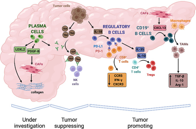
Pancreatic adenocarcinoma is highly infiltrated by B lymphocytes but the relevance of these immune cells in tumor development has been surprisingly overlooked until recently. Based on available evidence from other solid tumors, interaction between B lymphocytes and neoplastic cells is probably not uniformly stimulatory or inhibitory. Although presentation of tumor antigens to T cells and production of antitumor immunoglobulins might intuitively suggest a prominent tumor suppressive activity, specific subsets of B lymphocytes can secrete growth factors for neoplastic cells and immunosuppressive cytokines thus promoting escape from immunosurveillance and cancer progression. Because many of these mechanisms might also be implicated in the development of PDAC, and immune-modulation of B-cell activity is nowadays possible at different levels, determining the role of B-lymphocytes in this lethal cancer becomes of utmost importance to design novel therapeutic strategies. This review aims to discuss the emerging role of B cells in PDAC tumorigenesis, progression, and associated stromal reaction.

Keywords: B cells; B lymphocyte; PDAC - pancreatic ductal adenocarcinoma; cancer associated fibroblast (CAF); fibrosis; pancreatic adenocarcinoma.


Conflict of interest statement

The authors declare that the research was conducted in the absence of any commercial or financial relationships that could be construed as a potential conflict of interest.

Figures

Figure 1
Figure 1
Spatial organization of B lymphocytes in pancreatic adenocarcinoma. (A) Hematoxylin/Eosin stain and (B) immunofluorescence staining of a representative tissue section of PDAC showing an abundant infiltrate of B lymphocytes organized as either multiple lymphoid structures (asterisks) or spread within the neoplastic lesion and tumor stroma (arrowheads).
Figure 2
Figure 2
Evidence-based contribution of B lymphocytes to progression of pancreatic adenocarcinoma. B cells infiltrate PDAC in response to the release of local chemokines, such as CXCL13, and can contribute to both tumour promotion and tumour suppression through a variety of mechanisms. Tumour derived IL-18 can induce B lymphocytes with a regulatory phenotype that inhibit anti-tumour cytotoxic T cells responses. B-regulatory cells also secrete immune-modulatory cytokines such as IL-35 and IL-10 that induce T-regulatory cells (Tregs), stimulate tumour proliferation, and promote local angiogenesis. In addition, B-regulatory cells can recruit monocytes to tumour site and foster their differentiation into TGF-β and IL-10 producing M2 macrophages, thus further amplifying immune-evasive strategies and impairing cytotoxic responses. On the other hand, tumour-suppressive activity of B-cells in PDAC has not been clearly established but in-vitro studies suggest that it might depend on the production of autoantibodies against tumour associated antigen (TAA), engagement of natural killer cells (NK cells), and antigen dependent cell-mediated cytotoxicity (ADCC). Finally, plasma cells can sustain activation of the tumour stroma through the secretion of proliferative stimuli for cancer-associated fibroblasts (CAFs), such as PDGFB, and the production of enzymes that regulate extracellular matrix stiffness, such as LOXL2. Whether these properties ultimately exert tumour suppressive or tumour promoting effects is currently under investigation.

References

    1. Ryan DP, Hong TS, Bardeesy N. Pancreatic Adenocarcinoma. N Engl J Med (2014) 371:1039–49. doi: 10.1056/NEJMra1404198 - DOI - PubMed
    1. Rahib L, Fleshman JM, Matrisian LM, Berlin JD. Evaluation of Pancreatic Cancer Clinical Trials and Benchmarks for Clinicallymeaningful Future Trials: A Systematic Review. JAMA Oncol (2016) 2:1209–16. doi: 10.1001/jamaoncol.2016.0585 - DOI - PubMed
    1. Biankin AV, Kench JG, Dijkman FP, Biankin SA, Henshall SM. Molecular Pathogenesis of Precursor Lesions of Pancreatic Ductal Adenocarcinoma. Pathology (2003) 35:14–24. doi: 10.1080/003130202201472 - DOI - PubMed
    1. Hruban RH, Takaori K, Klimstra DS, Adsay NV, Albores-Saavedra J, Biankin AV, et al. . An Illustrated Consensus on the Classification of Pancreatic Intraepithelial Neoplasia and Intraductal Papillary Mucinous Neoplasms. Am J Surg Pathol (2004) 28:977–87. doi: 10.1097/01.pas.0000126675.59108.80 - DOI - PubMed
    1. Conroy T, Hammel P, Hebbar M, Ben Abdelghani M, Wei AC, Raoul J-L, et al. . FOLFIRINOX or Gemcitabine as Adjuvant Therapy for Pancreatic Cancer. N Engl J Med (2018) 379:2395–406. doi: 10.1056/nejmoa1809775 - DOI - PubMed

Publication types