

Blunt cerebrovascular injury of cervical and petrous ICA with stroke: A case study
- PMID: 35360003
- PMCID: PMC8961236
- DOI: 10.1016/j.tcr.2022.100637
Blunt cerebrovascular injury of cervical and petrous ICA with stroke: A case study
Abstract
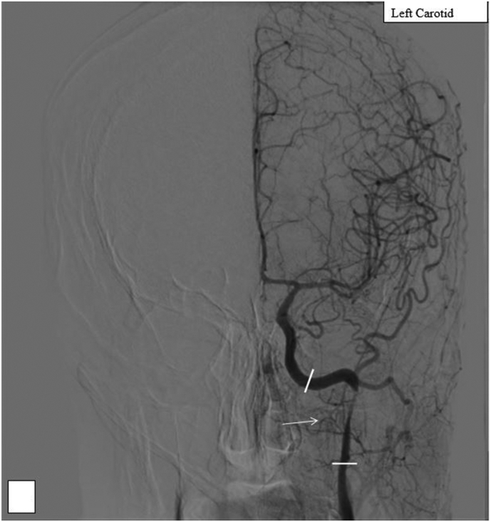
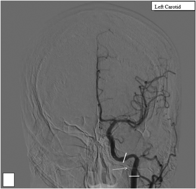
This case report documents treatment of a 32-year-old male patient after being extricated from an extensively damaged motor vehicle after a collision. On first presentation, the patient had a Glascow Coma Score of 14 and a pan-scan revealed a severely comminuted fracture of the proximal left femoral shaft, a fracture of the left patella, and multiple rib fractures. A neurological exam on a later evaluation showed global aphasia and sharp decline in movements of the right upper and lower extremities even with painful stimuli. A CT Angiography of the head and neck was obtained and revealed evidence of occlusion in the upper cervical segment and proximal petrous segment of the left internal carotid artery (ICA). Two 6x40mm precise stents were placed restoring normal intracranial flow to the patient's left ICA. The patient was discharged having sensation and motor function in all extremities and with resolved aphasia and no neurologic deficits.
Keywords: Blunt cerebrovascular injury; Neurologic deficits.
© 2022 The Authors.
Conflict of interest statement
No conflicts of interest to disclose by any of the authors, either financial or personal.
Figures
Similar articles
-
Successful stenting seven days after atherothrombotic occlusion of the intracranial internal carotid artery.J Endovasc Ther. 2006 Apr;13(2):254-9. doi: 10.1583/05-1742R.1. J Endovasc Ther. 2006. PMID: 16643082
-
Operative exposure and management of the petrous and upper cervical internal carotid artery.Neurosurgery. 1986 Dec;19(6):967-82. doi: 10.1227/00006123-198612000-00012. Neurosurgery. 1986. PMID: 3808244
-
Recanalisation of the internal carotid artery via the vasa vasorum after coil occlusion.Br J Radiol. 2011 Feb;84(998):e23-6. doi: 10.1259/bjr/31383692. Br J Radiol. 2011. PMID: 21257830 Free PMC article.
-
Blunt carotid artery injury.Emerg Med (Fremantle). 2002 Dec;14(4):412-21. doi: 10.1046/j.1442-2026.2002.00348.x. Emerg Med (Fremantle). 2002. PMID: 12534485 Review.
-
[A case of adult moyamoya disease showing fulminant clinical course associated with progression from unilateral to bilateral involvement].No Shinkei Geka. 1997 Jan;25(1):79-84. No Shinkei Geka. 1997. PMID: 8990473 Review. Japanese.
References
Publication types
LinkOut - more resources
Full Text Sources
Miscellaneous
