

The Impact of Intracoronary Imaging on PCI Outcomes in Cases Utilising Rotational Atherectomy: An Analysis of 8,417 Rotational Atherectomy Cases from the British Cardiovascular Intervention Society Database
- PMID: 35360091
- PMCID: PMC8941577
- DOI: 10.1155/2022/5879187
The Impact of Intracoronary Imaging on PCI Outcomes in Cases Utilising Rotational Atherectomy: An Analysis of 8,417 Rotational Atherectomy Cases from the British Cardiovascular Intervention Society Database
Abstract
Introduction: There is increasing evidence supporting the use of intracoronary imaging to optimize the outcomes of percutaneous coronary intervention (PCI). However, there are no studies examining the impact of imaging on PCI outcomes in cases utilising rotational atherectomy (RA-PCI). Our study examines the determinants and outcomes of using intracoronary imaging in RA-PCI cases including 12-month mortality.
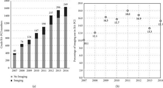
Methods: Using the British Cardiac Intervention Society database, data were analysed on all RA-PCI procedures in the UK between 2007 and 2014. Descriptive statistics and multivariate logistic regressions were used to examine baseline, procedural, and outcome associations with intravascular imaging.
Results: Intracoronary imaging was used in 1,279 out of 8,417 RA-PCI cases (15.2%). Baseline covariates associated with significantly more imaging use were number of stents used, smoking history, previous CABG, pressure wire use, proximal LAD disease, laser use, glycoprotein inhibitor use, cutting balloons, number of restenosis attempted, off-site surgery, and unprotected left main stem (uLMS) PCI. Adjusted rates of in-hospital major adverse cardiac/cerebrovascular events (IH-MACCE), its individual components (death, peri-procedural MI, stroke, and major bleed), or 12-month mortality were not significantly altered by the use of imaging in RA-PCI. However, subgroup analysis demonstrated a signal towards reduction in 12-month mortality in uLMS RA-PCI cases utilising intracoronary imaging (OR 0.67, 95% CI 0.44-1.03).
Conclusions: Intracoronary imaging use during RA-PCI is associated with higher risk of baseline and procedural characteristics. There were no differences observed in IH-MACCE or 12-month mortality with intracoronary imaging in RA-PCI.
Copyright © 2022 Majd B. Protty et al.
Conflict of interest statement
MM reports holding unrestricted educational grants from Boston Scientific, Terumo, and Abbott. All other authors report no conflicts of interest relevant to this work.
Figures


Similar articles
-
Rotational Atherectomy Complicated by Coronary Perforation Is Associated With Poor Outcomes: Analysis of 10,980 Cases From the British Cardiovascular Intervention Society Database.Cardiovasc Revasc Med. 2021 Jul;28:9-13. doi: 10.1016/j.carrev.2020.07.040. Epub 2020 Aug 2. Cardiovasc Revasc Med. 2021. PMID: 32888836
-
Excimer laser coronary atherectomy during complex PCI: An analysis of 1,471 laser cases from the British Cardiovascular Intervention Society database.Catheter Cardiovasc Interv. 2021 Apr 1;97(5):E653-E660. doi: 10.1002/ccd.29251. Epub 2020 Sep 18. Catheter Cardiovasc Interv. 2021. PMID: 32946132
-
Combined use of rotational and excimer lASER coronary atherectomy (RASER) during complex coronary angioplasty-An analysis of cases (2006-2016) from the British Cardiovascular Intervention Society database.Catheter Cardiovasc Interv. 2021 Jun 1;97(7):E911-E918. doi: 10.1002/ccd.29377. Epub 2020 Nov 17. Catheter Cardiovasc Interv. 2021. PMID: 33201601
-
Orbital atherectomy versus rotational atherectomy: A systematic review and meta-analysis.Int J Cardiol. 2020 Mar 15;303:16-21. doi: 10.1016/j.ijcard.2019.12.037. Epub 2019 Dec 17. Int J Cardiol. 2020. PMID: 31898984
-
Comparison of Rotational with Orbital Atherectomy During Percutaneous Coronary Intervention for Coronary Artery Calcification: A Systematic Review and Meta-Analysis.Cardiovasc Revasc Med. 2020 Apr;21(4):501-507. doi: 10.1016/j.carrev.2019.07.019. Epub 2019 Jul 22. Cardiovasc Revasc Med. 2020. PMID: 31377129
Cited by
-
Predictive value of the CHA₂DS₂-VASc-HSF score for slow flow during rotational atherectomy in patients with severe coronary artery calcification.BMC Cardiovasc Disord. 2025 Jul 4;25(1):469. doi: 10.1186/s12872-025-04931-1. BMC Cardiovasc Disord. 2025. PMID: 40615779 Free PMC article.
References
-
- Räber L., Mintz G. S., Koskinas K. C., et al. Clinical use of intracoronary imaging. Part 1: guidance and optimization of coronary interventions. An expert consensus document of the European Association of Percutaneous Cardiovascular Interventions. European Heart Journal . 2018;39(35):3281–3300. doi: 10.1093/eurheartj/ehy285. - DOI - PubMed
MeSH terms
LinkOut - more resources
Full Text Sources
Medical
Miscellaneous
