Review
. 2022 Mar 10:10:833163.
doi: 10.3389/fbioe.2022.833163. eCollection 2022.

Re-Endothelialization of Decellularized Liver Scaffolds: A Step for Bioengineered Liver Transplantation

Affiliations
Review

Re-Endothelialization of Decellularized Liver Scaffolds: A Step for Bioengineered Liver Transplantation

Kewei Li et al. Front Bioeng Biotechnol. .

Abstract

Bioengineered livers (BELs) are an attractive therapeutic alternative to address the donor organ shortage for liver transplantation. The goal of BELs technology aims at replacement or regeneration of the native human liver. A variety of approaches have been proposed for tissue engineering of transplantable livers; the current review will highlight the decellularization-recellularization approach to BELs. For example, vascular patency and appropriate cell distribution and expansion are critical components in the production of successful BELs. Proper solutions to these components of BELs have challenged its development. Several strategies, such as heparin immobilization, heparin-gelatin, REDV peptide, and anti-CD31 aptamer have been developed to extend the vascular patency of revascularized bioengineered livers (rBELs). Other novel methods have been developed to enhance cell seeding of parenchymal cells and to increase graft functionality during both bench and in vivo perfusion. These enhanced methods have been associated with up to 15 days of survival in large animal (porcine) models of heterotopic transplantation but have not yet permitted extended survival after implantation of BELs in the orthotopic position. This review will highlight both the remaining challenges and the potential for clinical application of functional bioengineered grafts.

Keywords: bioengineered livers (BELs); decellularization; heterotopic transplantation; liver transplantation; orthotopic transplantation; scaffolds.


Conflict of interest statement

JR was employed by Miromatrix Medical Inc. The remaining authors declare that the research was conducted in the absence of any commercial or financial relationships that could be construed as a potential conflict of interest. The reviewer JB declared a shared affiliation, with no collaboration, with one of the authors KL to the handling editor at the time of the review.

Figures

GRAPHICAL ABSTRACT
GRAPHICAL ABSTRACT
Native porcine livers are cannulated on the PV, IVC and SVC and perfused with Triton X-100 and SDS solutions to get a decellularized liver. Meanwhile, HUVECs are cultured in 2D tissue culture flasks, harvested and injected through the SHIVC of the decellularized liver, followed by the PV 24 h later to get endo-only BEL. After 12–14 days in media the seeded hepatocytes can be injected through the CBD to get co-seeded BEL.
FIGURE 1
FIGURE 1
Kaplan-Meier analysis of survival of the allograft group and the rBELs group. Survival rate at 24 h was significantly higher in the allograft group compared to rBELs group (4 of 4, 100% vs. 1 of 9, 11.1%, p = 0.01). The study endpoint was death or the presence of a death equivalent endpoint. Death equivalent endpoints were: absent graft blood flow, grade IV hepatic encephalopathy, and uncontrolled bleeding, and animals were euthanized.
FIGURE 2
FIGURE 2
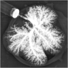
Portal venogram of Decell-recell liver graft demonstrating patency of vasculature and no evidence of thrombosis after 1-h in vivo perfusion.
FIGURE 3
FIGURE 3
(A). Decell-recell liver graft co-seeded with HUVEC endothelial cells and primary hepatocytes. (B). Light microscopy of co-seeded graft showing lobular structure after reseeding (H&E staining). (C). IHC microscopy of co-cultured graft showing mitotically active cells after reseeding (Ki67 staining).

References

    1. Agarwal T., Maiti T. K., Ghosh S. K. (2019). Decellularized Caprine Liver-Derived Biomimetic and Pro-angiogenic Scaffolds for Liver Tissue Engineering. Mater. Sci. Eng. C 98, 939–948. 10.1016/j.msec.2019.01.037 - DOI - PubMed
    1. Aji T., Dong J.-H., Shao Y.-M., Zhao J.-M., Li T., Tuxun T., et al. (2018). Ex Vivo liver Resection and Autotransplantation as Alternative to Allotransplantation for End-Stage Hepatic Alveolar Echinococcosis. J. Hepatol. 69 (5), 1037–1046. 10.1016/j.jhep.2018.07.006 - DOI - PubMed
    1. Anderson B. D., Nelson E. D., Joo D., Amiot B. P., Katane A. A., Mendenhall A., et al. (2021). Functional Characterization of a Bioengineered Liver after Heterotopic Implantation in Pigs. Commun. Biol. 4 (1), 1157. 10.1038/s42003-021-02665-2 - DOI - PMC - PubMed
    1. Ansari T., Southgate A., Obiri-Yeboa I., Jones L. G., Greco K., Olayanju A., et al. (2020). Development and Characterization of a Porcine Liver Scaffold. Stem Cell Dev. 29 (5), 314–326. 10.1089/scd.2019.0069 - DOI - PubMed
    1. Arulraj R., Neuberger J. (2011). Liver Transplantation: Filling the Gapbetween Supply and Demand. Clin. Med. 11 (2), 194–198. 10.7861/clinmedicine.11-2-194 - DOI - PMC - PubMed

LinkOut - more resources