Review
. 2022 Mar 14:10:850241.
doi: 10.3389/fbioe.2022.850241. eCollection 2022.

Nanomaterial-Based Immunocapture Platforms for the Recognition, Isolation, and Detection of Circulating Tumor Cells

Affiliations
Review

Nanomaterial-Based Immunocapture Platforms for the Recognition, Isolation, and Detection of Circulating Tumor Cells

Yichao Liu et al. Front Bioeng Biotechnol. .

Abstract

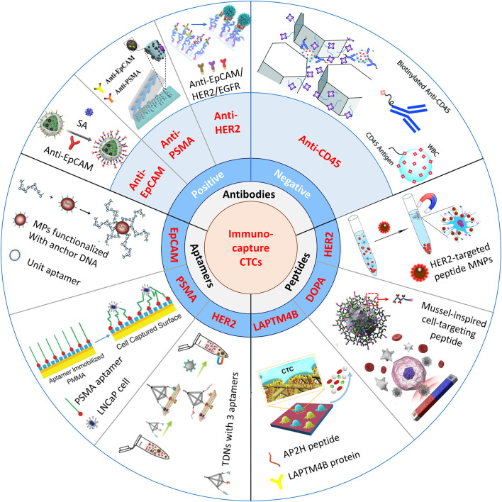
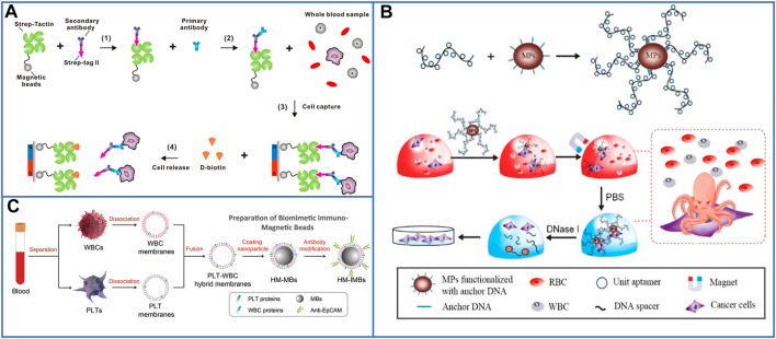
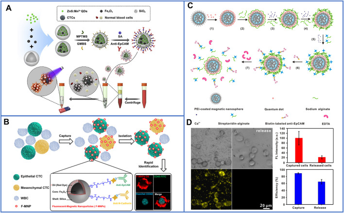
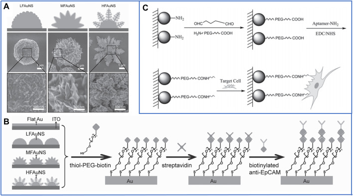
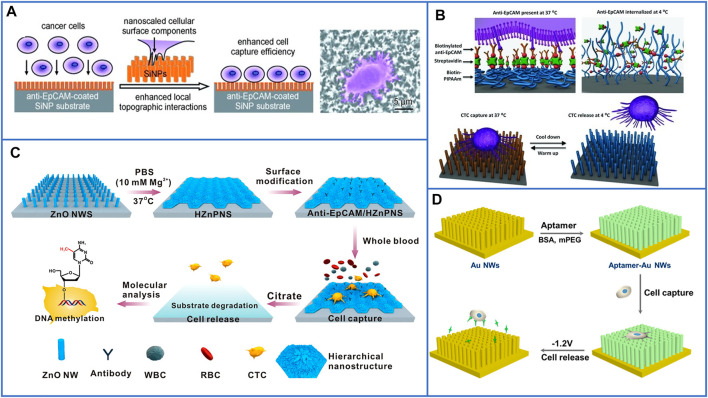
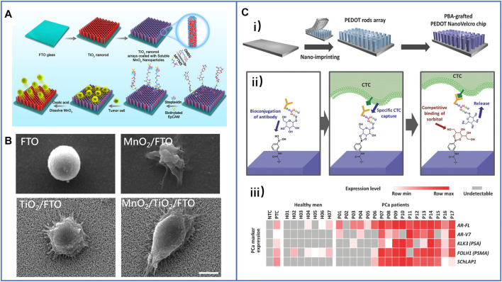
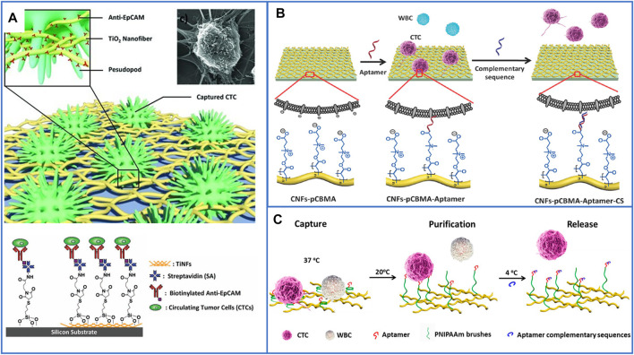
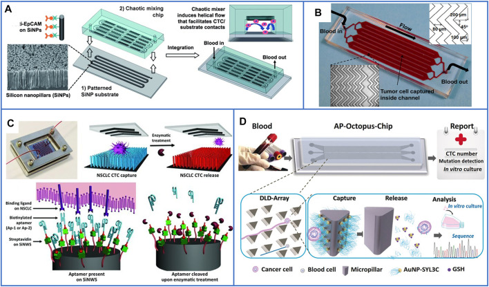
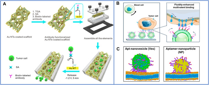
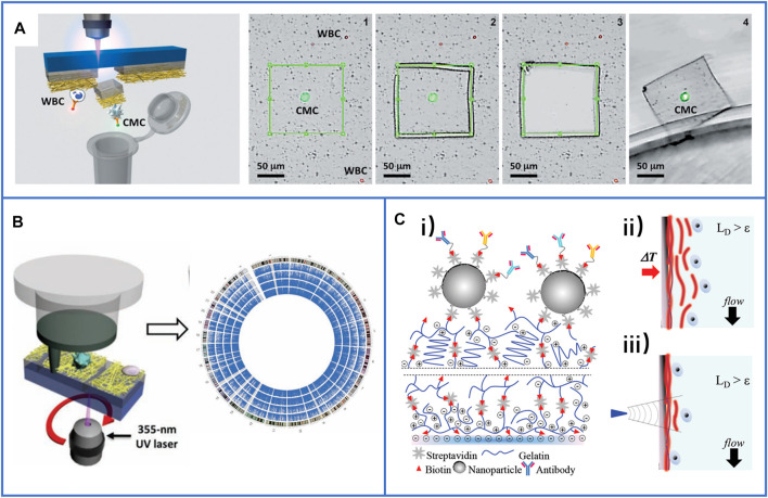
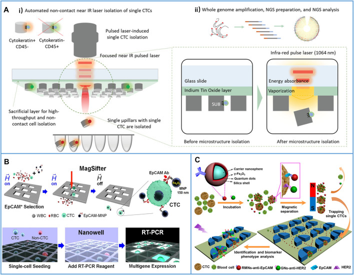
Circulating tumor cells (CTCs) are a type of cancer cells that circulate in the peripheral blood after breaking away from solid tumors and are essential for the establishment of distant metastasis. Up to 90% of cancer-related deaths are caused by metastatic cancer. As a new type of liquid biopsy, detecting and analyzing CTCs will provide insightful information for cancer diagnosis, especially the in-time disease status, which would avoid some flaws and limitations of invasive tissue biopsy. However, due to the extremely low levels of CTCs among a large number of hematologic cells, choosing immunocapture platforms for CTC detection and isolation will achieve good performance with high purity, selectivity, and viability. These properties are directly associated with precise downstream analysis of CTC profiling. Recently, inspired by the nanoscale interactions of cells in the tissue microenvironment, platforms based on nanomaterials have been widely explored to efficiently enrich and sensitively detect CTCs. In this review, various immunocapture platforms based on different nanomaterials for efficient isolation and sensitive detection of CTCs are outlined and discussed. First, the design principles of immunoaffinity nanomaterials are introduced in detail. Second, the immunocapture and release of platforms based on nanomaterials ranging from nanoparticles, nanostructured substrates, and immunoaffinity microfluidic chips are summarized. Third, recent advances in single-cell release and analysis of CTCs are introduced. Finally, some perspectives and challenges are provided in future trends of CTC studies.

Keywords: biological detection; circulating tumor cells; immunocapture platform; liquid biopsy; nanomaterials.


Conflict of interest statement

The authors declare that the research was conducted in the absence of any commercial or financial relationships that could be construed as a potential conflict of interest.

Figures

FIGURE 1
FIGURE 1
Schematic illustration of immunocapture platforms based on antibodies, peptides, and aptamers for CTC isolation. Positive antibodies: reproduced with permission from Cui et al. (2019), Copyright 2019, Elsevier. SA, streptavidin: reproduced with permission from Yin et al. (2018), Copyright 2018, American Chemical Society. PSMA, prostate-specific membrane antigen: reproduced with permission from Chen et al. (2019), Copyright 2019, John Wiley and Sons. HER2, human epidermal growth factor receptor 2. EGFR, epidermal growth factor receptor. Negative antibody: reproduced with permission from Chu et al. (2019), Copyright 2019, Royal Society of Chemistry. Peptides: reproduced with permission from Peng et al. (2017), Copyright 2017, American Chemical Society. MNPs, magnetic nanoparticles: reproduced with permission from Tian et al. (2019), Copyright 2019, American Chemical Society. DOPA, 3,4-dihydroxy-L-phenylalanine, a key functional amino acid in mussel adhesive proteins: reproduced with permission from Zhong et al. (2021), Copyright 2021, American Chemical Society. LAPTM4B, lysosomal protein transmembrane 4 β with extraordinarily high expression level in a majority of solid tumors. AP2H, a LAPTM4B-targeting peptide. Aptamers: reproduced with permission from Chen et al. (2019), Copyright 2019, American Chemical Society; reproduced with permission from Dharmasiri et al. (2009), Copyright 2009, John Wiley and Sons; reproduced with permission from Qin et al. (2020), Copyright 2020, John Wiley and Sons. TDNs, tetrahedral DNA nanostructures.
FIGURE 2
FIGURE 2
Magnetic nanoparticles to immunocapture CTCs. (A) Platform based on biotin-triggered decomposable immunomagnetic beads to efficiently capture and release viable CTCs: reproduced with permission from Lu et al. (2015), Copyright 2015, American Chemical Society. (B) A “NanoOctopus” platform based on long multimerized aptamer DNA strands to mimic octopus's tentacles for enhancing the sensitivity and specificity of immunomagnetic beads: reproduced with permission from Chen et al. (2019), Copyright 2019, American Chemical Society. (C) Schematic of PLT and WBC–hybrid membranes–modified immunomagnetic beads for highly enhancing the purity of the captured CTCs. Reproduced with permission from Rao et al. (2018), Copyright 2018, John Wiley and Sons.
FIGURE 3
FIGURE 3
Platforms based on skillfully designed fluorescent-magnetic nanoparticles for isolating and identifying CTCs. (A) Equipping ZnS:Mn2+ quantum dots and magnetic nanoparticles into hollow SiO2 nanospheres for capturing and conveniently identifying CTCs. Reproduced with permission from Cui et al. (2019), Copyright 2019, Elsevier. (B) Schematic of Fe3O4@SiO2 core-shell nanoparticles decorated with DiI dyes, and dual-antibody (anti-EpCAM and anti-N-cadherin) used for identifying CTCs from whole blood samples in only one-step processing. Reproduced with permission from Wang et al. (2019), Copyright 2019, American Chemical Society. (C) Schematic of fluorescent-MNPs coated by Ca2+-initiated alginate for capturing and identifying CTCs, and releasing viable CTCs. Reproduced with permission from Xie et al. (2014), Copyright 2014, American Chemical Society. (D) Microscopic images of isolated cancer cells and fluorescent identification by using Ca2+-initiated alginate-based fluorescent-MNPs, the two figures on the right of (D) show the capture yield and release yield of this platform. Reproduced with permission from Xie et al. (2014), Copyright 2014, American Chemical Society.
FIGURE 4
FIGURE 4
Fractal substrates based on nanoparticles to immunocapture CTCs. (A) Scanning electron microscope (SEM) images of three kinds of fractal gold nanostructures (FAuNSs) to capture and electrochemically release CTCs. Reproduced with permission from Zhang et al. (2013), Copyright 2013, John Wiley and Sons. (B) Schematic diagram of the chemically modified method on the surface of FAuNSs for capturing CTCs. Reproduced with permission from Zhang et al. (2013), Copyright 2013, John Wiley and Sons. (C) Fractal substrates based on chitosan nanoparticles and the chemically modified method for capturing CTCs. Reproduced with permission from Sun et al. (2015), Copyright 2015, John Wiley and Sons.
FIGURE 5
FIGURE 5
Fractal substrates based on nanopillars and nanowires to immunocapture CTCs. (A) The platform based on silicon-nanopillars array for highly enhancing the capture yield of CTCs. Reproduced with permission from Wang et al. (2014), Copyright 2009, John Wiley and Sons. (B) Schematic of silicon nanowire substrate coated with thermally responsive PIPAAm for capturing and releasing CTCs with high viability. Reproduced with permission from Hou et al. (2013), Copyright 2013, John Wiley and Sons. (C) A flowerlike substrate based on ZnO NWs coated with Mg2+ solutions to capture, rapidly release, and molecularly analyze viable CTCs. Reproduced with permission from Guo et al. (2016), Copyright 2016, American Chemical Society. (D) A platform based on AuNWs for capturing and releasing viable CTCs by using gently electrochemical method. Reproduced with permission from Zhai et al. (2017), Copyright 2017, American Chemical Society.
FIGURE 6
FIGURE 6
Fractal substrates based on nanorods to immunocapture CTCs. (A) The schematic process of TiO2 nanorod arrays coated with degradable MnO2 nanoparticles and conjugated with antibodies to capture and release CTCs. Reproduced with permission from Li et al. (2018), Copyright 2018, American Chemical Society. (B) SEM images of captured cancer cells on different substrates of FTO, MnO2/FTO, TiO2/FTO, and MnO2/TiO2/FTO. Scale bar: 5 μm. Reproduced with permission from Li et al. (2018), Copyright 2018, American Chemical Society. (C-i) Schematic illustration of PBA-grafted PEDOT NanoVelcro chip; (C-ii) the mechanism of conjugating antibody on PBA for CTCs capture and release; (C-iii) summary of RNA signature detection in blood samples of seven healthy men and CTCs of 17 PCa patients. Reproduced with permission from Shen et al. (2018), Copyright 2018, John Wiley and Sons.
FIGURE 7
FIGURE 7
Fractal substrates based on nanofibers to immunocapture CTCs. (A) The platform based on horizontal TiO2 electrospun nanofibers for highly enhancing capture yield of CTCs. Reproduced with permission (Zhang et al., 2012). Copyright 2012, John Wiley and Sons. (B) Schematic illustration of the design functional bio-interface based on chitosan nanofibers grafted with pCBMA brushes for highly enhancing the purity of captured CTCs and releasing cells. Reproduced with permission (Sun et al., 2016). Copyright 2016, John Wiley and Sons. (C) Schematic illustration of chitosan nanofibers coated with PNIPAAm for CTCs capture, purification and release. Reproduced with permission (Wang et al., 2017). Copyright 2017, American Chemical Society.
FIGURE 8
FIGURE 8
Microfluidic chips based on immunoaffinity nanomaterials for CTC isolation. (A) Schematic illustration of a microfluidic chip based on SiNP substrate and PDMS serpentine chaotic microchannel for capturing CTCs with high purity. Reproduced with permission from Wang et al. (2011), Copyright 2011, John Wiley and Sons. (B) A photo of microfluidic chip with herringbone mixers and AuNPs, the dimensions are shown in the top, right corner of the figure. Reproduced with permission from Sheng et al. (2013), Copyright 2013, American Chemical Society. (C) A microfluidic chip based on PDMS chaotic mixer and SiNWs substrate conjugated with two biotinylated DNA-aptamers for capturing and releasing CTCs. Reproduced with permission from Shen et al. (2013), Copyright 2013, John Wiley and Sons. (D) Working principle of the AP-Octopus-Chip based on AuNP-SYL3C–modified micropillar. Reproduced with permission from Song et al. (2019), Copyright 2019, John Wiley and Sons.
FIGURE 9
FIGURE 9
The microfluidic chips based on nanomaterials for highly enhancing the immunocapture efficiency of CTCs. (A) A 3D conductive scaffold microchip based on macroporous PDMS and immobilized gold nanotubes for effective capture and recovery of CTCs with high purity. Reproduced with permission from Cheng et al. (2021), Copyright 2021, American Chemical Society. (B) Working principle and diagram of the microfluidic chip based on natural nanovesicles for enhancing multivalent binding with cells. Reproduced with permission from Wu et al. (2021), Copyright 2020, American Chemical Society. (C) The simulations of the difference of aptamer scaffolds interacting with cell membranes and nanoparticles. Reproduced with permission from Wu et al. (2021), Copyright 2020, American Chemical Society.
FIGURE 10
FIGURE 10
Immunocapture-based platforms for single-cell isolation. (A) The process of the NanoVelcro chip with LMD technique to harvest single-CTCs. Reproduced with permission from Hou et al. (2013), Copyright 2013, John Wiley and Sons. (B) A polymer nanofiber-embedded microchip with LMD technique to get single-CTCs for the whole exome sequencing to understand the drug-resistant mechanisms of prostate cancer. Reproduced with permission from Zhao et al. (2013), Copyright 2013, John Wiley and Sons. (C-i) Schematic of a layer-by-layer nanocoating gelatin-based platform for the immunocapture of CTCs; (C-ii) schematic illustration of whole release of CTCs by raising the temperature to 37°C; (C-iii) schematic illustration of single-cell release by supplying local mechanical stress. Reproduced with permission from Reategui et al. (2015), Copyright 2015, John Wiley and Sons.
FIGURE 11
FIGURE 11
Immunocapture-based platforms for high-throughput single-cell isolation. (A) An optomechanically transferrable chip based on a near-IR light beam for single-CTC isolation for whole genome analysis. (A-i) Schematic of near-IR isolation of single-CTCs. (A-ii) Schematic of the downstream whole genome analysis of a single cell. Reproduced with permission from Kim et al. (2019), Copyright 2019, John Wiley and Sons. (B) Platform based on MagSifter and Nanowell devices for high-throughput single-CTC isolation and sensitive detection. Reproduced with permission from Park et al. (2016), Copyright 2016, National Academy of Sciences. (C) Micropillars-patterned microfluidic chip for convenient single-cell isolation and rapid in situ identification. Reproduced with permission from Wu et al. (2018), Copyright 2018, American Chemical Society.

Similar articles

Cited by

References

    1. Alix-Panabières C., Pantel K. (2013). Circulating Tumor Cells: Liquid Biopsy of Cancer. Clin. Chem. 59, 110–118. 10.1373/clinchem.2012.194258 - DOI - PubMed
    1. Arai F., Ng C., Maruyama H., Ichikawa A., El-Shimy H., Fukuda T. (2005). On Chip Single-Cell Separation and Immobilization Using Optical Tweezers and Thermosensitive Hydrogel. Lab. Chip 5, 1399–1403. 10.1039/B502546J - DOI - PubMed
    1. Ashworth T. (1869). A Case of Cancer in Which Cells Similar to Those in the Tumours Were Seen in the Blood after Death. Aust. Med. J. 14, 146.
    1. Bai L., Du Y., Peng J., Liu Y., Wang Y., Yang Y., et al. (2014). Peptide-based Isolation of Circulating Tumor Cells by Magnetic Nanoparticles. J. Mater. Chem. B 2, 4080–4088. 10.1039/c4tb00456f - DOI - PubMed
    1. Beech J. P., Holm S. H., Adolfsson K., Tegenfeldt J. O. (2012). Sorting Cells by Size, Shape and Deformability. Lab. Chip 12, 1048–1051. 10.1039/c2lc21083e - DOI - PubMed