

The Endonasal Endoscopic Approach to Different Sinonasal Fungal Balls
- PMID: 35360416
- PMCID: PMC8964197
- DOI: 10.1155/2022/6721896
The Endonasal Endoscopic Approach to Different Sinonasal Fungal Balls
Abstract
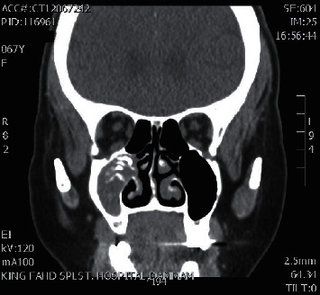
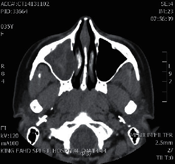
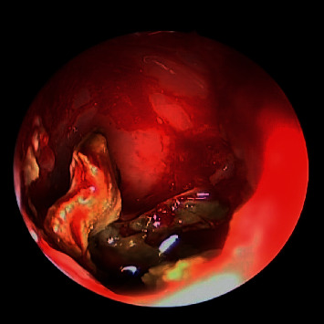
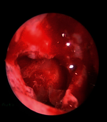
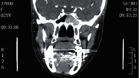
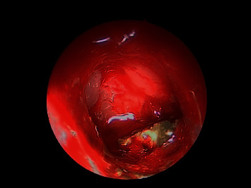
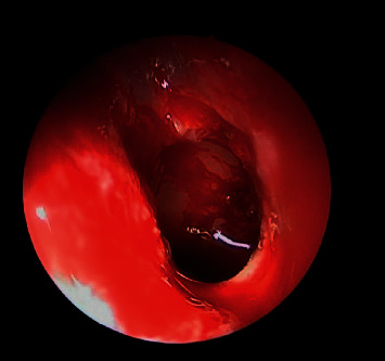
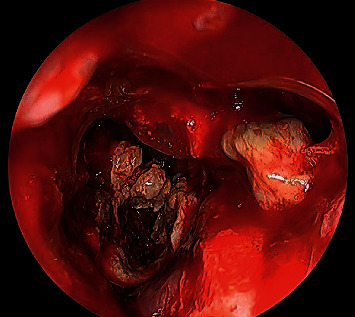
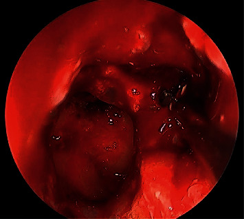
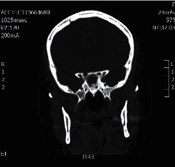
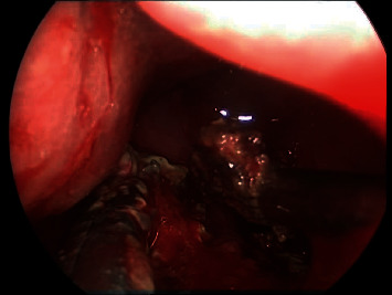
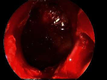
Background: Fungal ball sinusitis is a sinonasal fungus ball that usually affects immunocompetent adults with female predominance. The most affected sinus is the maxillary sinus. Aspergillus species is the most typically found fungus. Computed tomography (CT) scan is the gold standard tool in order to diagnose fungal ball sinusitis. The ultimate method for a fungal ball is functional endoscopic sinus surgery (FESS), which has a high success rate and a low morbidity rate.
Objective: This study aims to demonstrate the various clinical presentations of fungal ball sinusitis including isolated maxillary sinus, sphenoid sinus, simultaneous occurrence of maxillary and sphenoid fungal ball, and post endonasal endoscopic pituitary surgery fungal ball with various age groups. Also, this study aims to emphasize the importance of early diagnosis and treatment in such cases. Patients and Methods. A retrospective study that was carried in the otorhinolaryngology department of two hospitals: King Fahad Specialist Hospital and Qatif Central Hospital, Eastern Region, Saudi Arabia. The study was conducted on a total of 16 patients who were diagnosed with paranasal sinuses fungal ball in an 11-year period from January 2008 and November 2019.
Results: Out of 16 patients with paranasal sinuses fungal ball, 11 cases were female and 5 males, with age ranging between 16 and 46 years. Results showed eight isolated sphenoid (50%), six isolated maxillary fungal ball (38%), one simultaneous occurrence of the sphenoid and maxillary fungal ball (6%), and one post endonasal endoscopic pituitary surgery for pituitary adenoma (6%). CT scan was performed for all 16 cases which is the standard tool for the diagnosis of the fungal ball.
Conclusion: Fungal ball may present with variety of symptoms but most commonly with postnasal discharge (PND), headache, and facial pain. CT sinuses is the diagnostic radiological modality to confirm the diagnosis. The FESS functional endoscopic sinus surgery is the gold safe approach for patients with fungal ball to manage their symptoms, confirm the diagnosis, and removal of disease with no morbidities.
Copyright © 2022 Ali Almomen et al.
Conflict of interest statement
The authors declare that there are no conflicts of interest.
Figures
Similar articles
-
The simultaneous occurrence of a fungal ball in maxillary and sphenoid sinuses: a case report and review of the literature.J Surg Case Rep. 2025 Feb 28;2025(2):rjaf014. doi: 10.1093/jscr/rjaf014. eCollection 2025 Feb. J Surg Case Rep. 2025. PMID: 40040773 Free PMC article.
-
Endoscopic Findings of Fungal Ball in the Mucocele After Endonasal Transsphenoidal Surgery.Ear Nose Throat J. 2021 Mar;100(3):NP169-NP170. doi: 10.1177/0145561319867669. Epub 2019 Sep 25. Ear Nose Throat J. 2021. PMID: 31554429
-
Paranasal sinus fungus balls.Head Neck. 1997 Sep;19(6):481-6. doi: 10.1002/(sici)1097-0347(199709)19:6<481::aid-hed4>3.0.co;2-v. Head Neck. 1997. PMID: 9278755
-
Unusual Location of a Fungus Ball: The Concha Bullosa, a Review of the Literature.Allergy Rhinol (Providence). 2021 Aug 23;12:21526567211036146. doi: 10.1177/21526567211036146. eCollection 2021 Jan-Dec. Allergy Rhinol (Providence). 2021. PMID: 34457373 Free PMC article. Review.
-
Mucormycosis (Mucor fungus ball) of the maxillary sinus.Ear Nose Throat J. 2014 Oct-Nov;93(10-11):E18-22. Ear Nose Throat J. 2014. PMID: 25397383 Review.
References
-
- Dufour X., Kauffmann-Lacroix C., Ferrie J. C., Goujon J. M., Rodier M. H., Klossek J. M. Paranasal sinus fungus ball: epidemiology, clinical features and diagnosis. A retrospective analysis of 173 cases from a single medical center in France 1989–2002. Sabouraudia . 2006;44(1):61–67. - PubMed
LinkOut - more resources
Full Text Sources
