. 2022 Mar 14:13:810974.
doi: 10.3389/fgene.2022.810974. eCollection 2022.

Profile and Functional Prediction of Plasma Exosome-Derived CircRNAs From Acute Ischemic Stroke Patients

Affiliations

Profile and Functional Prediction of Plasma Exosome-Derived CircRNAs From Acute Ischemic Stroke Patients

Jie Yang et al. Front Genet. .

Abstract

Stroke is one of the major causes of death and long-term disability, of which acute ischemic stroke (AIS) is the most common type. Although circular RNA (circRNA) expression profiles of AIS patients have been reported to be significantly altered in blood and peripheral blood mononuclear cells, the role of exosome-containing circRNAs after AIS is still unknown. Plasma exosomes from 10 AIS patients and 10 controls were isolated, and through microarray and bioinformatics analysis, the profile and putative function of circRNAs in the plasma exosomes were studied. A total of 198 circRNAs were differentially quantified (|log2 fold change| ≥ 1.00, p < 0.05) between AIS patients and controls. The levels of 12 candidate circRNAs were verified by qRT-PCR, and the quantities of 10 of these circRNAs were consistent with the data of microarray. The functions of host genes of differentially quantified circRNAs, including RNA and protein process, focal adhesion, and leukocyte transendothelial migration, were associated with the development of AIS. As a miRNA sponge, differentially quantified circRNAs had the potential to regulate pathways related to AIS, like PI3K-Akt, AMPK, and chemokine pathways. Of 198 differentially quantified circRNAs, 96 circRNAs possessing a strong translational ability could affect cellular structure and activity, like focal adhesion, tight junction, and endocytosis. Most differentially quantified circRNAs were predicted to bind to EIF4A3 and AGO2-two RNA-binding proteins (RBPs)-and to play a role in AIS. Moreover, four of ten circRNAs with verified levels by qRT-PCR (hsa_circ_0112036, hsa_circ_0066867, hsa_circ_0093708, and hsa_circ_0041685) were predicted to participate in processes of AIS, including PI3K-Akt, AMPK, and chemokine pathways as well as endocytosis, and to be potentially useful as diagnostic biomarkers for AIS. In conclusion, plasma exosome-derived circRNAs were significantly differentially quantified between AIS patients and controls and participated in the occurrence and progression of AIS by sponging miRNA/RBPs or translating into proteins, indicating that circRNAs from plasma exosomes could be crucial molecules in the pathogenesis of AIS and promising candidates as diagnostic biomarkers and therapeutic targets for the condition.

Keywords: acute ischemic stroke; ceRNA; circRNA; plasma exosome; profile.


Conflict of interest statement

The authors declare that the research was conducted in the absence of any commercial or financial relationships that could be construed as a potential conflict of interest.

Figures

FIGURE 1
FIGURE 1
Identification and characterization of plasma exosomes. ( A ) Detection of CD63 and TSG101 as exosome markers by western blot. ( B,C ) Representative images of transmission electron microscopy. ( D,E ) Representative size of exosomes from controls and AIS patients, respectively.
FIGURE 2
FIGURE 2
CircRNA profiles of plasma exosomes from AIS patients (AIS) and controls (CON). (A) Heat map of circRNAs differentially quantified between AIS patients and controls. (B) Volcano plot of circRNA profiles between AIS patients and controls. Red dots represent significantly upregulated circRNAs (log2 fold change ≥ 1.00 and p < 0.05, n = 10, independent two-sample t-test), and green dots represent significantly downregulated circRNAs (log2 fold change ≤ −1.00 and p < 0.05, n = 10, independent two-sample t-test).
FIGURE 3
FIGURE 3
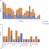
Characterization of circRNA profiles between AIS patients and controls. (A) Bar diagram of the distribution of differentially quantified circRNAs in different chromosomes. (B) Top 15 tissue-specific distributions of differentially quantified circRNAs.
FIGURE 4
FIGURE 4
GO annotations and KEGG pathway analyses for host genes of the differentially quantified circRNAs. (A) GO annotations. The GO analysis categorized the mRNAs into different groups under the theme of biological process, cellular component, and molecular function. The gene count for each GO category is listed on the left. (B) Top 10 KEGG pathways. The vertical axis indicates the pathway category, and the horizontal axis indicates the −log10 p-value of a pathway (Fisher’s exact test).
FIGURE 5
FIGURE 5
Validation and network diagram of differentially quantified circRNAs. ( A,B ) Relative fold changes of 12 differentially quantified circRNAs using qRT-PCR (n = 10). *, p < 0.05. ( C ) Network of circRNA-miRNA of 198 differentially quantified circRNAs. ( D ) Network of circRNA-miRNA-mRNA of the 10 differentially quantified circRNAs verified by qRT-PCR.
FIGURE 6
FIGURE 6
GO annotations and KEGG pathway analyses for target genes of the 10 differentially quantified circRNAs verified by qRT-PCR. (A) GO annotations. The GO analysis categorized the mRNAs into different groups under the theme of biological process, cellular component, and molecular function. The gene count for each GO category is listed on the left. (B) Top 10 KEGG pathways. The vertical axis indicates the pathway category, and the horizontal axis indicates the −log10 p-value of a pathway (Fisher’s exact test).
FIGURE 7
FIGURE 7
GO annotations and KEGG pathway analyses for proteins potentially coded by differentially quantified circRNAs. (A) Distribution of the coding probability of differentially quantified circRNAs. (B) GO annotations. The GO analysis categorized the genes into different groups under the theme of biological process, cellular component, and molecular function. The gene count for each GO category is listed on the left. (C) Top 10 KEGG pathways. The vertical axis indicates the pathway category, and the horizontal axis indicates the −log10 p-value of a pathway (Fisher’s exact test).
FIGURE 8
FIGURE 8
Distribution of top 15 RBPs predicted to bind to differentially quantified circRNAs.
FIGURE 9
FIGURE 9
ROC curves of four differentially quantified circRNAs verified by qRT-PCR, including ( A ) hsa_circ_0112036, ( B ) hsa_circ_0041685, ( C ) hsa_circ_0093708, and ( D ) hsa_circ_0066867 (n = 10). The red diagonal lines in the graphs represent the diagnostic threshold value of circRNAs for AIS (AUC = 0.500).

Similar articles

Cited by

References

    1. Abdelmohsen K., Panda A. C., Munk R., Grammatikakis I., Dudekula D. B., De S., et al. (2017). Identification of HuR Target Circular RNAs Uncovers Suppression of PABPN1 Translation by CircPABPN1. RNA Biol. 14 (3), 361–369. 10.1080/15476286.2017.1279788 - DOI - PMC - PubMed
    1. Abdullahi W., Tripathi D., Ronaldson P. T. (2018). Blood-brain Barrier Dysfunction in Ischemic Stroke: Targeting Tight Junctions and Transporters for Vascular protection. Am. J. Physiology-Cell Physiol. 315 (3), C343–c356. 10.1152/ajpcell.00095.2018 - DOI - PMC - PubMed
    1. Alberts M. J. (2017). Stroke Treatment with Intravenous Tissue-type Plasminogen Activator. Circulation 135 (2), 140–142. 10.1161/circulationaha.116.025724 - DOI - PubMed
    1. Ameli P. A., Ameli N. J., Gubernick D. M., Ansari S., Mohan S., Satriotomo I., et al. (2014). Role of Vasopressin and its Antagonism in Stroke Related Edema. J. Neurosci. Res. 92 (9), 1091–1099. 10.1002/jnr.23407 - DOI - PubMed
    1. Ashwal-Fluss R., Meyer M., Pamudurti N. R., Ivanov A., Bartok O., Hanan M., et al. (2014). circRNA Biogenesis Competes with Pre-mRNA Splicing. Mol. Cel 56 (1), 55–66. 10.1016/j.molcel.2014.08.019 - DOI - PubMed