

Treatment of calcaneonavicular coalition accompanied by an accessory anterolateral talar facet
- PMID: 35361102
- PMCID: PMC9057527
- DOI: 10.52312/jdrs.2022.496
Treatment of calcaneonavicular coalition accompanied by an accessory anterolateral talar facet
Abstract
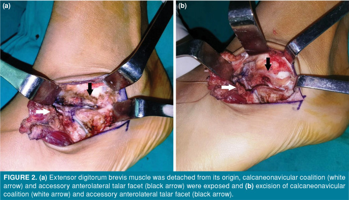
The association of accessory anterolateral talar facet (AALTF) and tarsal coalition has been reported recently. However, there is no report in the literature examining the clinical outcomes of operative treatment simultaneously addressing both AALTF and tarsal coalition. In this case series, we report the functional outcomes of operative treatment for both AALTF and calcaneonavicular coalition (CNC). Four male patients were admitted to our institution with foot pain. Radiographic examination revealed CNC and accompanying AALTF in all patients. Five feet of these four patients were operated simultaneously for AALTF and CNC. At the final follow-up, the mean Visual Analog Scale score was 1.7±2.4 (range, 0 to 5.5), the mean American Orthopedic Foot and Ankle Society score was 89.6±11.5 (range, 69 to 97), and the mean Foot Function Index was 15.4±19.1 (range, 0 to 43). In conclusion, simultaneous resection of CNC with AALTF seems to have good postoperative clinical outcomes. As AALTF can emerge along with CNC, every patient scheduled for CNC resection should be evaluated for AALTF.
Conflict of interest statement
Figures

References
-
- Sarrafian SK. Anatomy of the foot and ankle: descriptive, topographic, functional. 2nd ed. Philadelphia: Lippincott; 1993.
-
- Aydıngöz Ü, Topcuoğlu OM, Görmez A, Cankurtaran T, Topcuoğlu ED, Ergen FB. Accessory anterolateral talar facet in populations with and without symptoms: Prevalence and relevant associated ankle MRI findings. AJR Am J Roentgenol. 2016;207:846–851. - PubMed
MeSH terms
Supplementary concepts
LinkOut - more resources
Full Text Sources
Research Materials
