. 2022 May 11;42(19):3919-3930.
doi: 10.1523/JNEUROSCI.2416-21.2022. Epub 2022 Mar 31.

Synaptotagmins 1 and 7 Play Complementary Roles in Somatodendritic Dopamine Release

Affiliations

Synaptotagmins 1 and 7 Play Complementary Roles in Somatodendritic Dopamine Release

Takuya Hikima et al. J Neurosci. .

Abstract

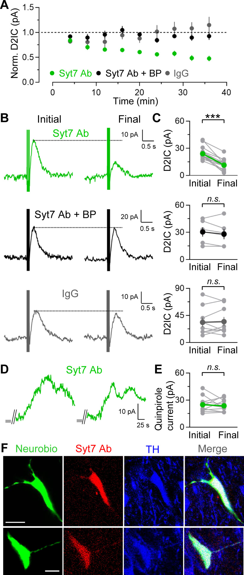
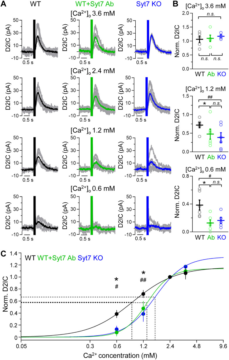
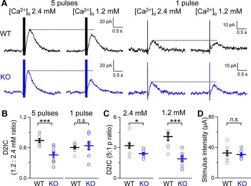
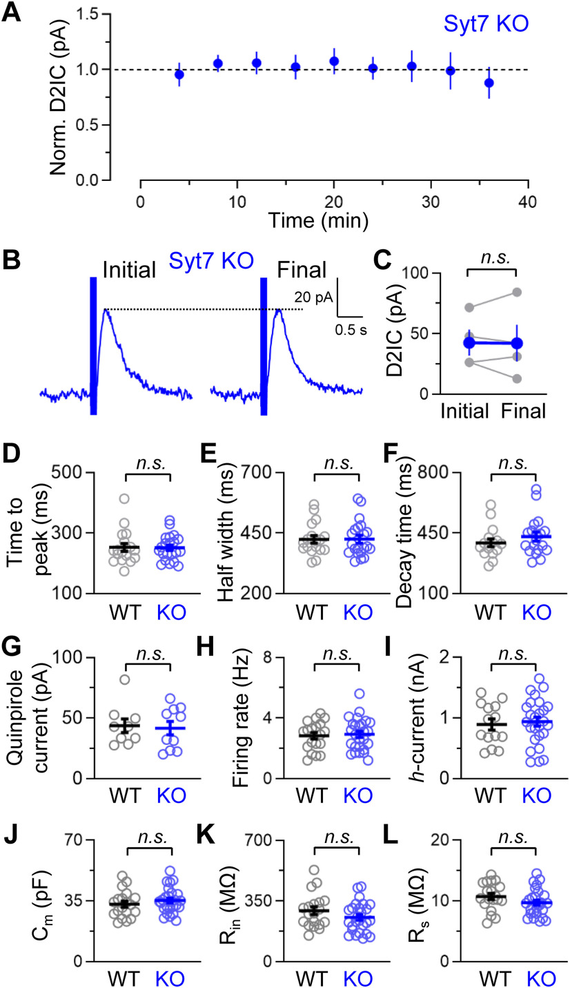
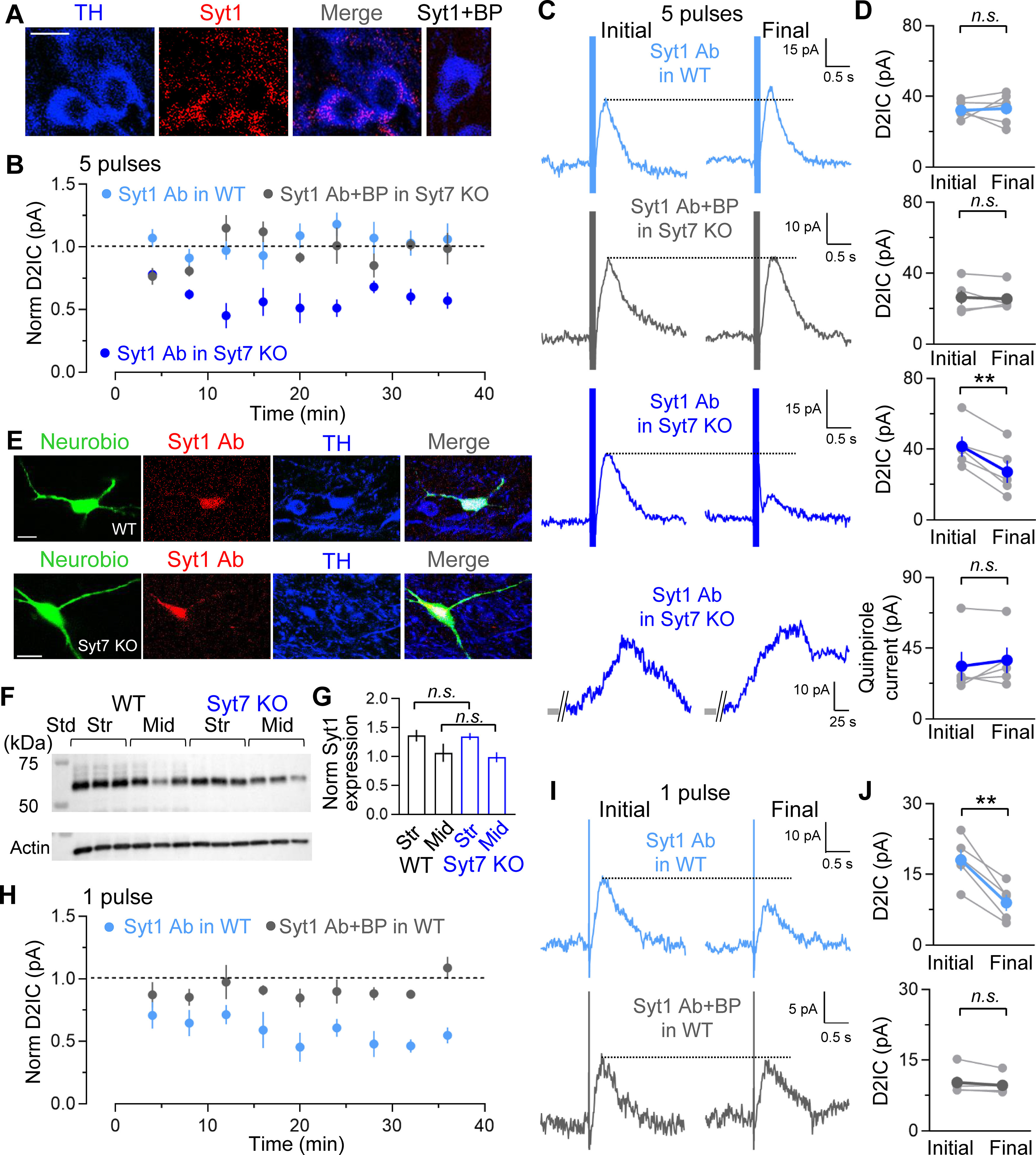
The molecular mechanisms underlying somatodendritic dopamine (DA) release remain unresolved, despite the passing of decades since its discovery. Our previous work showed robust release of somatodendritic DA in submillimolar extracellular Ca2+ concentration ([Ca2+]o). Here we tested the hypothesis that the high-affinity Ca2+ sensor synaptotagmin 7 (Syt7), is a key determinant of somatodendritic DA release and its Ca2+ dependence. Somatodendritic DA release from SNc DA neurons was assessed using whole-cell recording in midbrain slices from male and female mice to monitor evoked DA-dependent D2 receptor-mediated inhibitory currents (D2ICs). Single-cell application of an antibody to Syt7 (Syt7 Ab) decreased pulse train-evoked D2ICs, revealing a functional role for Syt7. The assessment of the Ca2+ dependence of pulse train-evoked D2ICs confirmed robust DA release in submillimolar [Ca2+]o in wild-type (WT) neurons, but loss of this sensitivity with intracellular Syt7 Ab or in Syt7 knock-out (KO) mice. In millimolar [Ca2+]o, pulse train-evoked D2ICs in Syt7 KOs showed a greater reduction in decreased [Ca2+]o than seen in WT mice; the effect on single pulse-evoked DA release, however, did not differ between genotypes. Single-cell application of a Syt1 Ab had no effect on train-evoked D2ICs in WT SNc DA neurons, but did cause a decrease in D2IC amplitude in Syt7 KOs, indicating a functional substitution of Syt1 for Syt7. In addition, Syt1 Ab decreased single pulse-evoked D2ICs in WT cells, indicating the involvement of Syt1 in tonic DA release. Thus, Syt7 and Syt1 play complementary roles in somatodendritic DA release from SNc DA neurons.SIGNIFICANCE STATEMENT The respective Ca2+ dependence of somatodendritic and axonal dopamine (DA) release differs, resulting in the persistence of somatodendritic DA release in submillimolar Ca2+ concentrations too low to support axonal release. We demonstrate that synaptotagmin7 (Syt7), a high-affinity Ca2+ sensor, underlies phasic somatodendritic DA release and its Ca2+ sensitivity in the substantia nigra pars compacta. In contrast, we found that synaptotagmin 1 (Syt1), the Ca2+ sensor underlying axonal DA release, plays a role in tonic, but not phasic, somatodendritic DA release in wild-type mice. However, Syt1 can facilitate phasic DA release after Syt7 deletion. Thus, we show that both Syt1 and Syt7 act as Ca2+ sensors subserving different aspects of somatodendritic DA release processes.

Keywords: D2 dopamine receptors; GIRK channels; autoreceptors; exocytosis; substantia nigra; synaptotagmin 1.


Figures

Figure 1.
Figure 1.
Syt7 immunoreactivity in SNc DA neurons and efficacy of Syt7 KO. A, Representative images showing immunostaining for Syt7 (red) and TH (blue) in the SNc of a coronal midbrain section in WT mice. Preadsorption of Syt7 with its BP resulted in loss of Syt7 immunostaining (Syt7+BP). Scale bar, 10 µm. B, Representative images showing immunostaining for Syt7 and TH in the SNc of a coronal midbrain section in Syt7 KO mice. Scale bar, 10 µm. C, Immunoblot of brain region extracts from WT or homozygous Syt7 KO mice probed with an affinity-purified antibody against the Syt7 confirms specificity of the Syt7 Ab and the efficacy of the KO. Cx, Cortex; Cer, cerebellum; Hip, hippocampus; Str, striatum; Mid, midbrain; Std, protein molecular mass standard.
Figure 2.
Figure 2.
Intracellular application of Syt7 Ab decreases D2IC amplitude. A, Average time course of changes in the amplitude of phasic D2ICs (five pulses, 40 Hz) with Syt7 Ab, Syt7 Ab+BP, or IgG in the recording pipette. Intracellular application of Syt7 Ab alone decreased amplitude; coinfusion of the BP for Syt7 Ab nullified this effect. B, Representative D2ICs after establishing whole-cell recording (initial) and at the end of the recording period (final). C, Quantification of D2IC amplitude when recorded with Syt7, Syt7 Ab+BP, or IgG (Syt7 Ab: final, 48 ± 5% of initial, n = 15; Syt7 Ab+BP: final, 91 ± 6% of initial, n = 6; p = 0.26; IgG: final, 111 ± 14% of initial, n = 9; p = 0.74, paired t test). D, Representative D2-dependent currents evoked by brief superfusion of quinpirole (250 nm, 15 s). Quinpirole was applied immediately after establishing whole-cell recording (initial) and at the end of the recording period (final) with Syt7 Ab in the pipette. E, Quantification of quinpirole-evoked D2 currents in the presence of Syt7 (Syt7 Ab: final, 99 ± 8% of initial, n = 12; p = 0.66, paired t test). F, Representative immunostaining of Syt7 Ab and TH in the SNc in two horizontal slices after whole-cell recording. Neurobiotin 488 was used to identify patch-clamped cells and was introduced together with Syt7 Ab via the recording pipette (n = 6 slices). Scale bars, 10 µm. n.s., Not significant. ***p < 0.0001.
Figure 3.
Figure 3.
Intracellular application of Syt7 Ab in SNc DA neurons delayed the time to peak of evoked D2ICs compared with controls, but did not alter other D2IC parameters. A, Time to peak (Syt7 Ab: initial, 204 ± 10 ms; final, 265 ± 13 ms, n =7; p = 0.004; Syt7 Ab+BP: initial, 196 ± 7 ms; final, 208 ± 2 ms, n = 5; p = 0.15; IgG: initial, 278 ± 24 ms; final, 296 ± 29 ms, n =7; p = 0.35). B, Half-width (duration at 50% peak amplitude): Syt7 Ab: initial, 359 ± 9 ms; final, 369 ± 15 ms; p = 0.56; Syt7 Ab+BP: initial, 310 ± 20 ms; final, 318 ± 20 ms; p = 0.22; IgG: initial, 410 ± 30 ms; final, 421 ± 31 ms; p = 0.32). C, Decay time constant (Syt7 Ab: initial, 396 ± 25 ms; final, 412 ± 37 ms; p = 0.73; Syt7 Ab+BP: initial, 304 ± 24 ms; final, 268 ± 20 ms; p = 0.69; IgG: initial, 405 ± 31 ms; final, 364 ± 41 ms; p = 0.44, paired t test). n.s., Not significant. ***p < 0.001.
Figure 4.
Figure 4.
Influence of Syt7 on the Ca2+ dependence of somatodendritic DA release. A, Evoked D2ICs in the SNc in varying [Ca2+]o. Averaged records are shown in color, and single D2IC traces are shown in gray. In control WT SNc DA neurons, robust D2ICs were seen in 1.2 and 0.6 mm [Ca2+]o. However, D2ICs were minimal in low [Ca2+]o when recorded with intracellular Syt7 Ab or in SNc DA neurons from Syt7 KO mice. B, D2IC amplitudes from WT, WT+Syt7 Ab, and Syt7 KO SNc DA neurons normalized to D2IC amplitude in 2.4 mm [Ca2+]o for each cell (3.6 mm: WT vs Ab, p = 0.79; WT vs KO, p = 0.75; Ab vs KO, p = 0.79; 1.2 mm: WT vs Ab, p = 0.042; WT vs KO, p = 0.0054; Ab vs KO, p = 0.38; 0.6 mm: WT vs Ab, p = 0.011; WT vs KO, p = 0.042; Ab vs KO, p = 0.62; n = 4–6/condition, two-way ANOVA with Holm–Sidak multiple-comparisons test). C, Ca2+ dependence of somatodendritic DA release in SNc, normalized to peak amplitude in 2.4 mm [Ca2+]o for each condition. Solid lines show the Hill fit for WT (black), Syt7 Ab in WT (green), and Syt7 KO (blue). The calculated Hill coefficients were 1.8 for WT, 3.3 for WT+Syt7 Ab, and 2.6 for Syt7 KO. The x-axis for each Hill plot was extended to 9.6 mm [Ca2+]o to permit extrapolation of the Ca2+ dependence to an approximate maximal level for each region. These plots were also used to calculate the [Ca2+]o at which DA release is half-maximal (EC50), indicated by dashed lines: WT EC50 was 0.9 mm [Ca2+]o; WT+Syt7 Ab was 1.3 mm [Ca2+]o; and Syt7 KO was 1.6 mm [Ca2+]o (n = 4–6 for each data point, two-way ANOVA with Holm–Sidak multiple-comparisons test). n.s., Nonsignificant. *p < 0.05, WT vs Syt7 Ab; #p < 0.05, WT vs Syt7 KO; ##p < 0.01, for WT vs Syt7 KO.
Figure 5.
Figure 5.
The influence of Syt7 on phasic versus tonic somatodendritic DA release. A, Representative D2ICs elicited by one-pulse (tonic) or five-pulse (phasic) stimulation at 40 Hz in 2.4 or 1.2 mm [Ca2+]o in SNc DA neurons from WT and Syt7 KO mice. B, Ratio of D2IC amplitude in 1.2 mm [Ca2+]o to 2.4 mm [Ca2+]o as an index of the Ca2+ dependence in WT and Syt7 KO mice. The effect of decreasing Ca2+ on five-pulse evoked D2ICs was significantly greater in Syt7 KO mice versus WT, whereas no genotypic difference was seen with one-pulse evoked D2ICs (phasic D2ICs: WT, 1.2 mm 0.76 ± 0.03 of 2.4 mm, n = 9; Syt7 KO, 1.2 mm 0.46 ± 0.05 of 2.4 mm, n = 8; p = 0.0004; tonic D2ICs: WT, 1.2 mm 0.60 ± 0.03 of 2.4 mm, n = 9; Syt7 KO: 1.2 mm 0.64 ± 0.07 of 2.4 mm, n = 8; p = 0.65, unpaired t test). C, Ratio of five-pulse to one-pulse (5:1 p ratio) evoked D2IC amplitude in a given SNc DA neuron shows that Syt7 amplifies the contrast between phasic and tonic somatodendritic DA release (2.4 mm: WT, 3.2 ± 0.2, n = 9; Syt7 KO, 2.5 ± 0.2, n = 8; p = 0.018; 1.2 mm: WT, 4.1 ± 0.4, n = 9; Syt7 KO, 1.9 ± 0.2, n = 8; p = 0.0004, unpaired t test). D, Stimulation intensities used to evoked D2ICs did not differ between WT and Syt7 KO SNc DA neurons (WT: 33.0 ± 0.3 μA, n = 9; Syt7 KO: 31.0 ± 0.2 μA, n = 8; p = 0.61, unpaired t test). n.s., Not significant. *p < 0.05, ***p < 0.001.
Figure 6.
Figure 6.
Characteristics of persistent D2ICs in SNc DA neurons from Syt7 KO mice. A, Stability of phasic D2ICs in SNc DA neurons from Syt7 KO mice recorded in 2.4 mm [Ca2+]o. B, Representative D2ICs after establishing whole-cell recording (initial) and at the end of the recording period (final). C, Quantification of D2IC amplitude (final: 93 ± 14% of initial, n = 4; p = 0.95, unpaired t test). D, Time to peak (WT: 253 ± 13 ms, n = 18; KO: 251 ± 9 ms, n = 21; p = 0.93, unpaired t test). E, Half-width (WT: 424 ± 15 ms, n = 18; KO: 425 ± 16 ms, n = 21; p = 0.98, unpaired t test). F, Decay time constant (WT: 394 ± 21 ms, n = 16; KO: 429 ± 24 ms, n =19; p = 0.32, unpaired t test). G, Quinpirole-evoked D2 currents (WT: 43.7 ± 5.3 pA, n = 9; Syt7 KO: 41.6 ± 5.2 pA, n = 10; p = 0.79, paired t test). H, Spontaneous activity (WT: 2.8 ± 0.2 Hz, n = 19; Syt7 KO: 2.9 ± 0.2 Hz, n = 26; p = 0.75, unpaired t test). I, h-current (WT: 0.89 ± 0.09 nA, n = 14; Syt7 KO: 0.93 ± 0.07 nA, n = 26; p = 0.72, unpaired t test). J, Cell capacitance (WT: 33.1 ± 1.7 pF, n = 19; Syt7 KO: 35.1 ± 1.4 pF, n = 26; p = 0.35, unpaired t test). K, Input resistance (WT: 293.9 ± 22.1 MΩ, n = 19; Syt7 KO: 255.8 ± 16.5 MΩ, n = 26; p = 0.17, unpaired t test). L, Series resistance (WT: 8.4 ± 0.8 MΩ, n = 19; Syt7 KO: 9.7 ± 0.4 MΩ, n = 26; p = 0.14, unpaired t test). n.s., Not significant.
Figure 7.
Figure 7.
Intracellular application of Syt1 Ab decreases D2IC amplitude in Syt7 KO mice, but not in WT mice. A, Representative images showing immunostaining for Syt1 (red) and TH (blue) in the SNc of a coronal section. Preadsorption of Syt1 with its BP resulted in the loss of Syt1 immunostaining (Syt1+BP). Scale bar, 10 µm. B, Average time course of changes in the amplitude of burst-evoked D2ICs (five pulses, 40 Hz) in 2.4 mm [Ca2+]o with Syt1 Ab in the recording pipette. Intracellular application of Syt1 Ab deceased amplitude in Syt7 KO mice, but not in WT mice. C, Representative initial and final D2ICs with Syt1 Ab in the pipette in WT (light blue) or Syt7 KO (blue) SNc DA neurons. Below, Representative D2 currents evoked by brief superfusion of quinpirole (250 nm, 15 s) with Syt1 Ab in the pipette. D, Quantification of D2IC amplitude with Syt1 Ab in the pipette (Syt1 Ab in WT: final, 105 ± 11% of initial, n = 6, p = 0.78; Syt1 Ab+BP in WT: final, 100 ± 7% of initial, n = 5, p = 0.71; Syt1 Ab in Syt7 KO: final, 63 ± 7% of initial, n = 5; paired t test). Quantification of quinpirole-evoked D2 currents in the presence of Syt1 Ab (Syt1 Ab: final, 119 ± 15% of initial, n = 5; p = 0.76, paired t test). E, Representative immunostaining of Syt1 Ab and TH in the SNc in a horizontal slice after whole-cell recording. Neurobiotin 488 was used to identify the patch-clamped cell and was introduced with Syt1 Ab via the recording pipette (n = 6 slices in three WT mice; n =7 slices in Syt7 KO mice). Scale bar, 10 µm. F, Immunoblots for Syt1 in striatum (Str) and midbrain (Mid) from WT and Syt7 KO mice (n = 3 mice/genotype). G, Quantification of Syt1 protein in WT and Syt7 KO midbrain and striatal tissue. H, Average time course of changes in the amplitude of one-pulse evoked D2ICs in 2.4 mm [Ca2+]o with Syt1 Ab in the recording pipette. Intracellular application of Syt1 Ab decreased amplitude in WT mice, but not with Syt1 Ab+BP. I, Representative initial and final D2ICs in WT SNc DA neurons recorded with Syt1 Ab or with Syt1 Ab+BP in the pipette. J, Quantification of D2IC amplitude with single-cell application of Syt1 Ab or Syt1+BP (Syt1 Ab: final, 50 ± 5% of initial, n = 5; Syt1 Ab+BP: final, 95 ± 2% of initial, n = 5; p = 0.15, paired t test). Scale bars: A, G, 20 µm. n.s., Not significant. **p < 0.01.

Similar articles

Cited by

References

    1. Augustine GJ, Charlton MP, Smith SJ (1985) Calcium entry and transmitter release at voltage-clamped nerve terminals of squid. J Physiol 367:163–181. 10.1113/jphysiol.1985.sp015819 - DOI - PMC - PubMed
    1. Bacaj T, Wu D, Yang X, Morishita W, Zhou P, Xu W, Malenka RC, Südhof TC (2013) Synaptotagmin-1 and -7 trigger synchronous and asynchronous phases of neurotransmitter release. Neuron 80:947–959. 10.1016/j.neuron.2013.10.026 - DOI - PMC - PubMed
    1. Baker K, Gordon SL, Grozeva D, van Kogelenberg M, Roberts NY, Pike M, Blair E, Hurles ME, Chong WK, Baldeweg T, Kurian MA, Boyd SG, Cousin MA, Raymond FL (2015) Identification of a human synaptotagmin-1 mutation that perturbs synaptic vesicle cycling. J Clin Invest 125:1670–1678. 10.1172/JCI79765 - DOI - PMC - PubMed
    1. Baker K, et al. . (2018) SYT1-associated neurodevelopmental disorder: a case series. Brain 141:2576–2591. 10.1093/brain/awy209 - DOI - PMC - PubMed
    1. Banerjee A, Lee J, Nemcova P, Liu C, Kaeser PS (2020) Synaptotagmin-1 is the Ca2+ sensor for fast striatal dopamine release. Elife 9:e58359. 10.7554/eLife.58359 - DOI - PMC - PubMed