. 2022 Mar 31;12(1):5496.
doi: 10.1038/s41598-022-09410-7.

SARS-CoV-2 spike protein induces cognitive deficit and anxiety-like behavior in mouse via non-cell autonomous hippocampal neuronal death

Affiliations

SARS-CoV-2 spike protein induces cognitive deficit and anxiety-like behavior in mouse via non-cell autonomous hippocampal neuronal death

Junyoung Oh et al. Sci Rep. .

Abstract

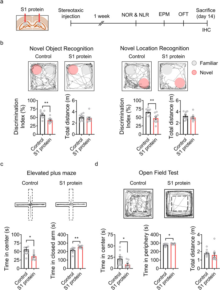
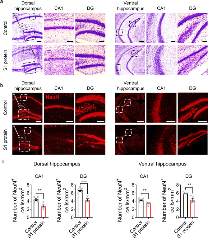
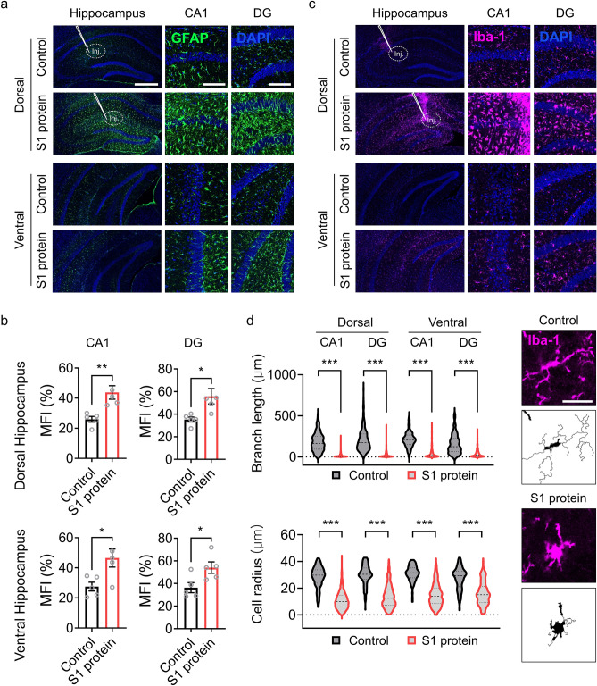
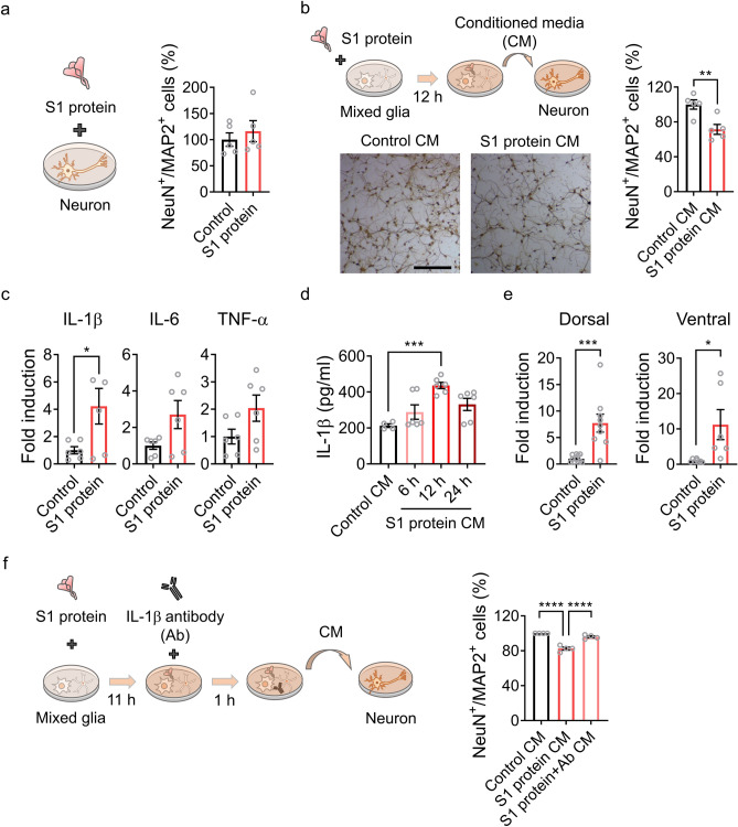
Severe acute respiratory syndrome coronavirus 2 (SARS-CoV-2) infection is accompanied by chronic neurological sequelae such as cognitive decline and mood disorder, but the underlying mechanisms have not yet been elucidated. We explored the possibility that the brain-infiltrating SARS-CoV-2 spike protein contributes to the development of neurological symptoms observed in COVID-19 patients in this study. Our behavioral study showed that administration of SARS-CoV-2 spike protein S1 subunit (S1 protein) to mouse hippocampus induced cognitive deficit and anxiety-like behavior in vivo. These neurological symptoms were accompanied by neuronal cell death in the dorsal and ventral hippocampus as well as glial cell activation. Interestingly, the S1 protein did not directly induce hippocampal cell death in vitro. Rather, it exerted neurotoxicity via glial cell activation, partially through interleukin-1β induction. In conclusion, our data suggest a novel pathogenic mechanism for the COVID-19-associated neurological symptoms that involves glia activation and non-cell autonomous hippocampal neuronal death by the brain-infiltrating S1 protein.


Conflict of interest statement

The authors declare no competing interests.

Figures

Figure 1
Figure 1
Administration of SARS-CoV-2 S1 protein induces cognitive deficit and anxiety-like behavior in mice. (a) Experimental design of S1 administration and behavioral tests. S1 protein (n = 10) or saline (Control, n = 10) was administered to C57BL/6 mice at 8 weeks of age, and behavioral tests were started 1 week after the administration. (b) Cognitive deficits were assessed using novel object recognition (NOR, left) or novel location recognition (NLR, right) tests and are presented as discrimination index percentages. Locomotor activity was measured by the total distance moved by mice in the chamber within a test session (bottom). The exploration time of a novel object or location divided by total exploration time was presented as the discrimination index of the novel object or location. The pink circle indicates a novel object or novel location. Anxiety-like behavior was assessed using the elevated plus maze test (EPM, panel c) and the open field test (OFT, panel d). (c) EPM trace of Control and S1 protein-injected groups (upper); the time spent in the open arm (dotted) vs. the closed arm (solid) in the EPM (bottom). (d) The center and periphery zones in the OFT are shown (upper). The time spent in the center vs. periphery zones in the OFT was examined (bottom). All representative navigations on the behavior test for a 5-min period are presented for the mice. The data are presented as the mean ± s.e.m. Statistical results are for unpaired t-tests. IHC, immunohistochemistry. ** p < 0.01, * p < 0.05.
Figure 2
Figure 2
SARS-CoV-2 S1 protein induces hippocampal neuronal death in CA1 and DG areas. (a-b). Hippocampal neurons in mice injected with S1 protein (n = 5) or vehicle (Control, n = 5) were visualized by cresyl violet staining (purple, scale bars = 200, 50, and 200 µm for left, middle, and right of dorsal area; 500 µm for left and 125 µm for middle and right of ventral area) and immunostaining with NeuN antibody (red, scale bars = 200, 50, and 100 µm for left, middle, and right of dorsal area; 500 µm for left and 125 µm for middle and right of ventral area). Hippocampal slices were prepared 14 days post-injection. (c) NeuN-positive cells in the CA1 and DG regions of the dorsal and ventral hippocampus were manually counted using ImageJ software (Wayne Rasband, National Institutes of Health, USA) in a blind manner. Data are presented as the mean ± s.e.m. Statistical results are for unpaired t-tests. **** p < 0.0001, ** p < 0.01, * p < 0.05.
Figure 3
Figure 3
S1 protein induces astrocyte and microglia activation in the hippocampus. (a, c) Representative images of immunohistochemistry with GFAP and Iba-1 antibodies show activation of astrocytes (GFAP+, green) and microglia (Iba-1+, purple) after stereotaxic S1 protein administration (n = 5, scale bar = 500 for left and 250 µm for middle and right). (b) Mean fluorescence intensity (MFI) of GFAP+ signal measured in CA1 and DG regions of dorsal hippocampus (upper) and ventral hippocampus (bottom). (d) Microglia activation was characterized by morphological parameters such as total branch length (upper left) and radius of cell area (bottom left). Representative pictures of microglia in the saline (Control)- or S1 protein-injected mice (right, scale bar = 25 µm). Data are presented as the `mean ± s.e.m. Statistical results are for unpaired t-tests. *** p < 0.001.
Figure 4
Figure 4
S1 protein induces non-cell autonomous hippocampal cell death via IL-1β induction in glial cells. (a) Primary hippocampal neurons were stimulated with S1 protein (5 µg/ml) for 12 h, and NeuN+ and MAP2+ cells were manually and blindly counted. The relative cell numbers in S1 protein-treated samples compared to the control samples are shown (right). (b) A schematic diagram (upper) showing primary hippocampal neuron stimulation by S1 protein-activated glia-conditioned media (CM). After 12 h, neuronal cells were counted, and relative cell numbers in the CM-treated samples are presented (right). Representative images of hippocampal neurons with or without CM treatment for 12 h (bottom). (c) Primary glia were stimulated with or without S1 protein for 12 h. Total RNA was prepared, and transcripts of proinflammatory cytokines were measured by real-time RT-PCR analysis. The gene expression levels were normalized against GAPDH in each target and are presented as fold induction. (d) IL-1β protein levels in the CM of primary glia with or without S1 treatment were measured by ELISA. (e) IL-1β expression in the hippocampus in vivo 6 h after administration of S1 protein. IL-1β transcript levels in the dorsal and ventral hippocampus were separately examined. (f) An experimental scheme testing the neurotoxicity of glial cell-derived IL-1β in CM using IL-1β-neutralizing antibody. Pretreatment with IL-1β antibody for 1 h rescues hippocampal neuronal death by CM. Data are presented as the mean ± s.e.m. Statistical results are from unpaired t-tests. **** p < 0.0001, *** p < 0.001, * p < 0.05.

References

    1. Lu R, et al. Genomic characterisation and epidemiology of 2019 novel coronavirus: Implications for virus origins and receptor binding. Lancet. 2020;395:565–574. doi: 10.1016/S0140-6736(20)30251-8. - DOI - PMC - PubMed
    1. Cucinotta D, Vanelli M. WHO Declares COVID-19 a Pandemic. Acta Biomed. 2020;91:157–160. - PMC - PubMed
    1. Hampshire, A. et al. Cognitive deficits in people who have recovered from COVID-19. E Clin.Med., 101044 (2021). - PMC - PubMed
    1. Xiong J, et al. Impact of COVID-19 pandemic on mental health in the general population: A systematic review. J. Affect Disord. 2020;277:55–64. doi: 10.1016/j.jad.2020.08.001. - DOI - PMC - PubMed
    1. Choi, E.P.H., Hui, B.P.H. & Wan, E.Y.F. Depression and anxiety in Hong Kong during COVID-19. Int. J. Environ. Res. Public Health17 (2020). - PMC - PubMed

Publication types