. 2022 Apr 1;78(Pt 4):472-482.
doi: 10.1107/S2059798322001802. Epub 2022 Mar 11.

Molecular insight into 2-phosphoglycolate activation of the phosphatase activity of bisphosphoglycerate mutase

Affiliations

Molecular insight into 2-phosphoglycolate activation of the phosphatase activity of bisphosphoglycerate mutase

Anfal S Aljahdali et al. Acta Crystallogr D Struct Biol. .

Abstract

Bisphosphoglycerate mutase (BPGM) is an erythrocyte-specific multifunctional enzyme that is responsible for the regulation of 2,3-bisphosphoglycerate (2,3-BPG) in red blood cells through its synthase and phosphatase activities; the latter enzymatic function is stimulated by the endogenous activator 2-phosphoglycolate (2-PG). 2,3-BPG is a natural allosteric effector of hemoglobin (Hb) that is responsible for decreasing the affinity of Hb for oxygen to facilitate tissue oxygenation. Here, crystal structures of BPGM with 2-PG in the presence and absence of 3-phosphoglycerate are reported at 2.25 and 2.48 Å resolution, respectively. Structure analysis revealed a new binding site for 2-PG at the dimer interface for the first time, in addition to the expected active-site binding. Also, conformational non-equivalence of the two active sites was observed as one of the sites was found in an open conformation, with the residues at the active-site entrance, including Arg100, Arg116 and Arg117, and the C-terminus disordered. The kinetic result is consistent with the binding of 2-PG to an allosteric or noncatalytic site as well as the active site. This study paves the way for the rational targeting of BPGM for therapeutic purposes, especially for the treatment of sickle cell disease.

Keywords: 2,3-bisphosphoglycerate; 2-phosphoglycolate; X-ray crystallography; bisphosphoglycerate mutase; kinetics; phosphatases; synthases.


Figures

Figure 1
Figure 1
Crystal structure of the ternary BPGM·3-PGA·2-PG complex. (a) Overall structure of BPGM·3-PGA·2-PG (ribbons) with 3-PGA (sticks) bound at the active site of monomer A and 2-­PG (sticks) bound at the active site of monomer B and at the dimer interface. (b) Electron-density map of the active site of monomer A with bound 3-PGA (2F oF c map contoured at the 0.8σ level). (c) Electron-density map of the active site of monomer B with bound 2-PG (2F oF c map contoured at the 0.8σ level). (d) Detailed interactions between the active-site residues of monomer A (cyan sticks) and 3-PGA (yellow sticks). (e) Detailed interactions between the active-site residues of monomer B (pink sticks) and 2-PG (green sticks). (f) Electron-density map of the dimer interface residues with bound 2-PG (2F o − F c map contoured at the 0.8σ level). (g) Detailed interactions between the dimer interface residues (cyan sticks for monomer A and pink sticks for monomer B) and 2-PG (green sticks).
Figure 2
Figure 2
Crystal structure of the binary BPGM·2-PG complex. (a) Overall structure of BPGM·2-PG (ribbons) with bound 2-PG (sticks) at the active sites of monomers A and B and at the dimer interface. (b) Electron-density map of the active site of monomer A with bound 2-PG (2F oF c map contoured at the 0.8σ level). (c) Electron-density map of the active site of monomer B with bound 2-PG (2F oF c map contoured at the 0.8σ level). (d) Detailed interactions between the active-site residues of monomer A (cyan sticks) and 2-PG (green sticks). (e) Detailed interactions between the active-site residues of monomer B (pink sticks) and 2-PG (green sticks). (f) Electron-density map of the dimer interface with bound 2-PG (2F oF c map contoured at the 0.8σ level). (g) Detailed interactions between the dimer interface residues (cyan sticks for monomer A and pink sticks for monomer B) and 2-PG (green sticks).
Figure 3
Figure 3
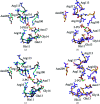
Structural comparison of the ternary BPGM·3-PGA·2-PG, binary BPGM·2-PG and unliganded BPGM (PDB entry 3nfy) structures. (a) Superposition of the active site of monomer A of BPGM·3-PGA·2-PG (cyan) and PDB entry 3nfy (gray). (b) Superposition of the active site of monomer B of BPGM·3-PGA·2-PG (pink) and PDB entry 3nfy (yellow). (c) Superposition of the active site of monomer A of BPGM·2-PG (cyan) and PDB entry 3nfy (gray). (d) Superposition of the active site of monomer B of BPGM·2-PG (violet) and PDB entry 3nfy (yellow)
Figure 4
Figure 4
(a) The relationship between V max and the 2-PG concentration. (b) The relationship between the apparent K m and the 2-PG concentration.

Similar articles

Cited by

References

    1. Afonine, P. V., Grosse-Kunstleve, R. W., Echols, N., Headd, J. J., Moriarty, N. W., Mustyakimov, M., Terwilliger, T. C., Urzhumtsev, A., Zwart, P. H. & Adams, P. D. (2012). Acta Cryst. D68, 352–367. - PMC - PubMed
    1. Benesch, R. & Benesch, R. E. (1967). Biochem. Biophys. Res. Commun. 26, 162–167. - PubMed
    1. Benesch, R. & Benesch, R. E. (1969). Nature, 221, 618–622. - PubMed
    1. Bunn, H. F. (1997). N. Engl. J. Med. 337, 762–769. - PubMed
    1. Bunn, H. F. & Forget, B. G. (1986). Hemoglobin: Molecular, Genetic and Clinical Aspects. Philadelphia: W. B. Saunders.

MeSH terms