. 2022 Apr 1;78(Pt 4):494-508.
doi: 10.1107/S2059798322001772. Epub 2022 Mar 11.

Crystal structure of the putative cell-wall lipoglycan biosynthesis protein LmcA from Mycobacterium smegmatis

Affiliations

Crystal structure of the putative cell-wall lipoglycan biosynthesis protein LmcA from Mycobacterium smegmatis

Onisha Patel et al. Acta Crystallogr D Struct Biol. .

Abstract

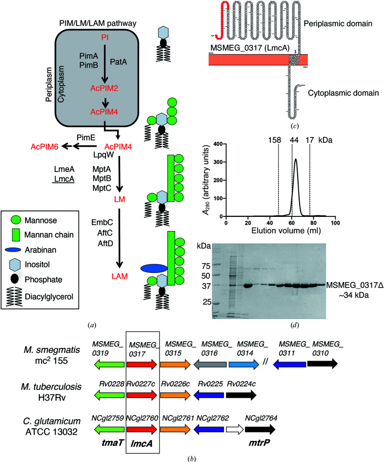
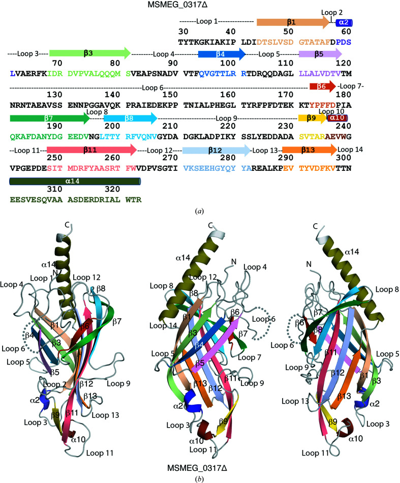
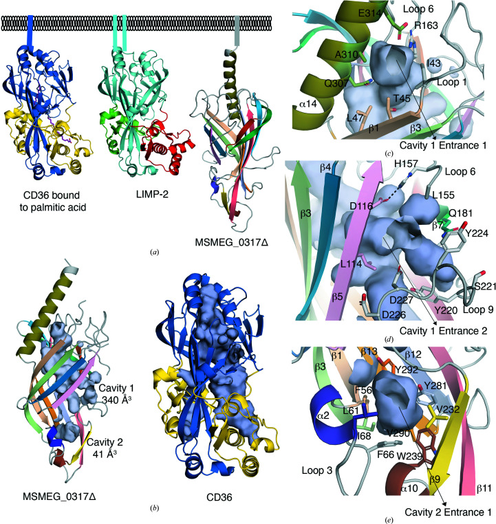
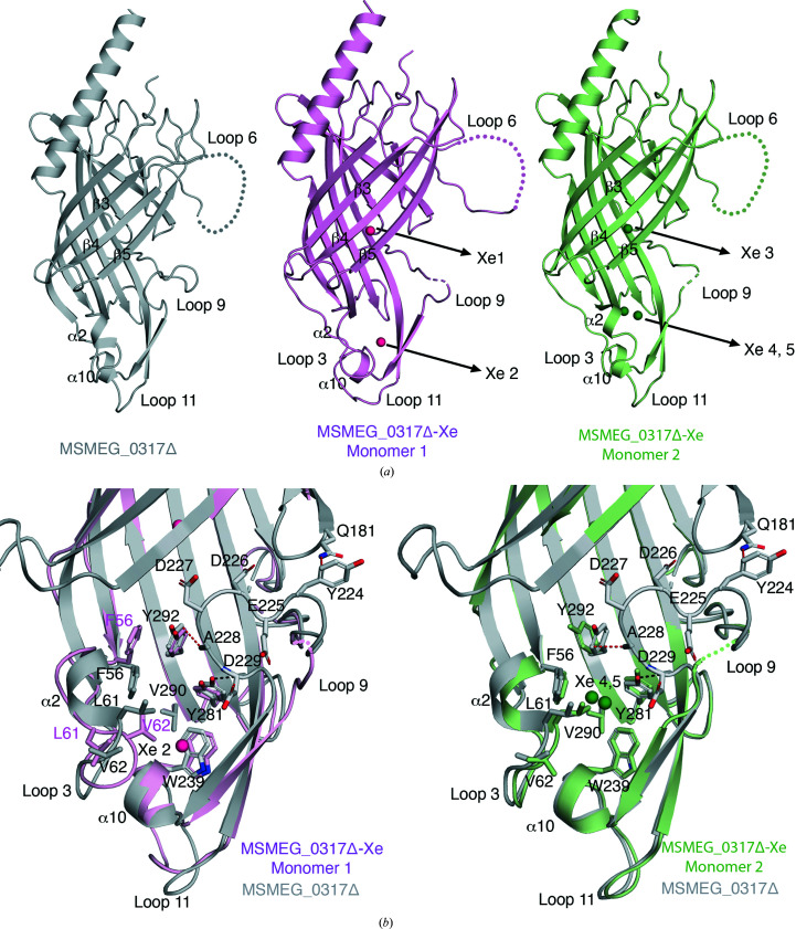
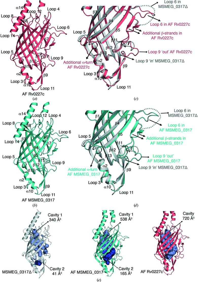
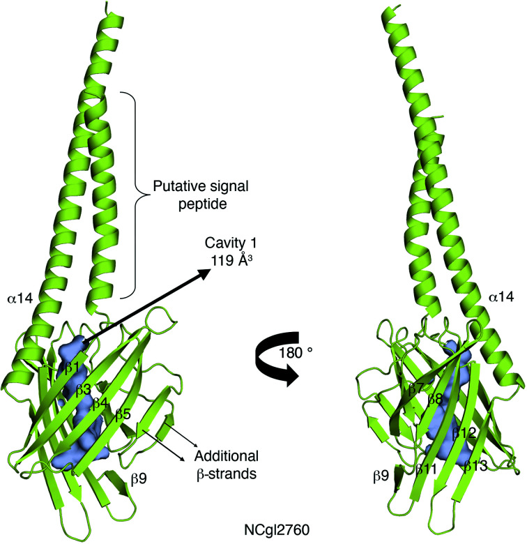
The bacterial genus Mycobacterium includes important pathogens, most notably M. tuberculosis, which infects one-quarter of the entire human population, resulting in around 1.4 million deaths from tuberculosis each year. Mycobacteria, and the closely related corynebacteria, synthesize a class of abundant glycolipids, the phosphatidyl-myo-inositol mannosides (PIMs). PIMs serve as membrane anchors for hyperglycosylated species, lipomannan (LM) and lipoarabinomannan (LAM), which are surface-exposed and modulate the host immune response. Previously, in studies using the model species Corynebacterium glutamicum, NCgl2760 was identified as a novel membrane protein that is required for the synthesis of full-length LM and LAM. Here, the first crystal structure of its ortholog in Mycobacterium smegmatis, MSMEG_0317, is reported at 1.8 Å resolution. The structure revealed an elongated β-barrel fold enclosing two distinct cavities and one α-helix extending away from the β-barrel core, resembling a `cone with a flake' arrangement. Through xenon derivatization and structural comparison with AlphaFold2-derived predictions of the M. tuberculosis homolog Rv0227c, structural elements were identified that may undergo conformational changes to switch from `closed' to `open' conformations, allowing cavity access. An AlphaFold2-derived NCgl2760 model predicted a smaller β-barrel core with an enclosed central cavity, suggesting that all three proteins, which were collectively termed LmcA, may have a common mechanism of ligand binding through these cavities. These findings provide new structural insights into the biosynthetic pathway for a family of surface lipoglycans with important roles in mycobacterial pathogenesis.

Keywords: MSMEG_0317; Mycobacterium smegmatis; Mycobacterium tuberculosis; lipoarabinomannan; lipomannan.


Figures

Figure 1
Figure 1
(a) The PIM/LM/LAM biosynthetic pathway of mycobacteria. Early steps of PIM synthesis are performed by the cytoplasmic enzymes PimA (Korduláková et al., 2002 ▸), PimB (Guerin et al., 2009; Lea-Smith et al., 2008 ▸) and PatA (Korduláková et al., 2003 ▸) to produce AcPIM2 from phosphatidylinositol (PI). Further mannosylation yields AcPIM4, which is transported to the periplasm and can be processed by the mannosyltransferase PimE (Morita et al., 2006 ▸) to form AcPIM6, an end product, or channelled into a parallel pathway for LM and LAM synthesis by the lipoprotein LpqW (Crellin et al., 2008; Kovacevic et al., 2006; Marland et al., 2006 ▸). LM/LAM synthesis is catalysed by the PPM-dependent mannosyltransferases MptB, MptA and MptC (Kaur et al., 2006, 2007; Mishra et al., 2007, 2008; Mishra, Krumbach et al., 2011 ▸). A phospholipid-binding protein, LmeA (Rahlwes et al., 2017 ▸), is involved in maintaining MptA under stress conditions (Rahlwes et al., 2020 ▸). The focus of the current study, LmcA (underlined), also functions at the MptA step in C. glutamicum (Cashmore et al., 2017 ▸). (b) The MSMEG_0317 genetic locus. The MSMEG_0317 gene is encoded within a locus that is highly conserved in Corynebacterineae. Likely orthologous genes in the three species are shown using the same colour. Previously studied genes are tmaT (Yamaryo-Botte et al., 2015 ▸) and mtrP (Rainczuk et al., 2020 ▸), both with roles in cell-wall mycolic acid transport, and the LM/LAM biosynthesis gene NCgl2760 (Cashmore et al., 2017 ▸), while the remaining genes are uncharacterized. The focus of the current study is boxed. (c) Predicted membrane topology of MSMEG_0317. Following cleavage of the putative signal peptide (red), the mature protein is proposed to comprise a large periplasmic N-­terminal domain, a single transmembrane domain and a small cytoplasmic tail. (d) The elution profile of MSMEG_0317Δ on a HiLoad 16/60 Superdex 75 gel-filtration column suggesting a monomeric protein (top) and SDS–PAGE analysis of the eluted MSMEG_0317Δ (∼34 kDa) (bottom). The molecular-weight markers used for calibration are bovine γ-globulin (158 kDa), chicken ovalbumin (44 kDa) and equine myoglobin (17 kDa). See also Supplementary Fig. S1.
Figure 2
Figure 2
The amino-acid sequence and the crystal structure of the periplasmic domain of MSMEG_0317Δ. (a) The sequence of MSMEG_0317Δ showing secondary-structure elements derived from the crystal structure of MSMEG_0317Δ. (b) The crystal structure of MSMEG_0317Δ in different views. The secondary-structure elements are colour-coded. The disordered loop 6 is shown by dotted lines. See also Supplementary Figs. S2–S4.
Figure 3
Figure 3
Structural homology and surface representation of the enclosed cavities in MSMEG_0317Δ. (a) Comparison of the MSMEG_0317Δ fold with the CD36 superfamily of scavenger receptor proteins, including the human lysosomal integral membrane protein 2 (LIMP-2) and CD36, a fatty-acid transporter, which show an extended asymmetric β-barrel core. (b) Comparison of the MSMEG_0317Δ enclosed cavities with the CD36 cavity which binds palmitic acid. (c) Close-up of MSMEG_0317Δ cavity 1 entrance 1. (d) Close-up of MSMEG_0317Δ cavity 1 entrance 2. (e) Close-up of MSMEG_0317Δ cavity 2. Hydrogen-bond and salt-bridge interactions are shown as black dashed lines. See also Supplementary Fig. S5.
Figure 4
Figure 4
The crystal structure of xenon-derivatized MSMEG_0317Δ (referred to as MSMEG_0317Δ-Xe). (a) Comparison of the crystal structures of monomer 1 and monomer 2 of MSMEG_0317Δ with MSMEG_0317Δ-Xe. The positions of the five Xe atoms (Xe 1 to Xe 5) in monomers 1 and 2 of MSMEG_0317Δ-Xe are highlighted. The disordered loops 6 and 9 are shown as dotted lines. (b) Overlay of the crystal structure of MSMEG_0317Δ with MSMEG_0317Δ-Xe and close-up view of the base of the β-barrel core to highlight conformational flexibility near the region of loop 3, the α2 turn, loop 11 and loop 9. Loop 9 adopts a closed conformation in MSMEG_0317Δ, while in MSMEG_0317Δ-Xe loop 9 is disordered (dotted line). Hydrogen-bond interactions are shown as black dashed lines and van der Waals interactions are shown as red dashed lines. See also Supplementary Fig. S6.
Figure 5
Figure 5
AlphaFold2-derived predictions of MSMEG_0317 (AF MSMEG_0317) and Rv0227c (AF Rv0227c). (a) Structure of AF Rv0227c. (b) Structure of AF MSMEG_0317Δ. (c) Overlay of the crystal structure of MSMEG_0317Δ with AF Rv0227c and a close-up view of the base of the β-barrel core. Loop 6 in AF Rv0227c is resolved and this loop contains two additional β-strands. Loop 9 in AF Rv0227c adopts an ‘out’ or ‘open’ conformation, in contrast to loop 9 in the MSMEG_0317Δ crystal structure, which adopts an ‘in’ or ‘closed’ conformation. (d) Overlay of the crystal structure of MSMEG_0317Δ with AF MSMEG_0317 and a close-up view of the base of the β-barrel core. Loop 6 in AF MSMEG_0317 is resolved in a similar position as in AF Rv0227c, including the additional two β-strands. Like AF Rv0227c, loop 9 in AF MSMEG_0317 adopts an ‘out’ or ‘open’ conformation. (e) Comparison of the enclosed cavities of the MSMEG_0317Δ, AF Rv0227c and AF MSMEG_0317 models. See also Supplementary Fig. S7.
Figure 6
Figure 6
AlphaFold2-derived prediction of NCgl2760 (AF NCgl2760). AF NCgl2760 adopts a smaller β-­barrel core compared with MSMEG_0317 and Rv0227c; however, the central cavity is still a conserved feature. Note that the N-terminal helix may represent a signal peptide. See also Supplementary Figs. S8 and S9.

References

    1. Alderwick, L. J., Lloyd, G. S., Ghadbane, H., May, J. W., Bhatt, A., Eggeling, L., Fütterer, K. & Besra, G. S. (2011). PLoS Pathog. 7, e1001299. - PMC - PubMed
    1. Aragão, D., Aishima, J., Cherukuvada, H., Clarken, R., Clift, M., Cowieson, N. P., Ericsson, D. J., Gee, C. L., Macedo, S., Mudie, N., Panjikar, S., Price, J. R., Riboldi-Tunnicliffe, A., Rostan, R., Williamson, R. & Caradoc-Davies, T. T. (2018). J. Synchrotron Rad. 25, 885–891. - PMC - PubMed
    1. Birch, H. L., Alderwick, L. J., Bhatt, A., Rittmann, D., Krumbach, K., Singh, A., Bai, Y., Lowary, T. L., Eggeling, L. & Besra, G. S. (2008). Mol. Microbiol. 69, 1191–1206. - PMC - PubMed
    1. Brennan, P. J. & Nikaido, H. (1995). Annu. Rev. Biochem. 64, 29–63. - PubMed
    1. Bricogne, G., Blanc, E., Brandl, M., Flensburg, C., Keller, P., Paciorek, W., Roversi, P., Sharff, A., Smart, O. S., Vonrhein, C. & Womack, T. O. (2017). BUSTER. Global Phasing Ltd, Cambridge, United Kingdom.

MeSH terms

LinkOut - more resources