

Facilitating standardized COVID-19 suspicion prediction based on computed tomography radiomics in a multi-demographic setting
- PMID: 35362751
- PMCID: PMC8973680
- DOI: 10.1007/s00330-022-08730-6
Facilitating standardized COVID-19 suspicion prediction based on computed tomography radiomics in a multi-demographic setting
Abstract
Objective: To develop an automatic COVID-19 Reporting and Data System (CO-RADS)-based classification in a multi-demographic setting.
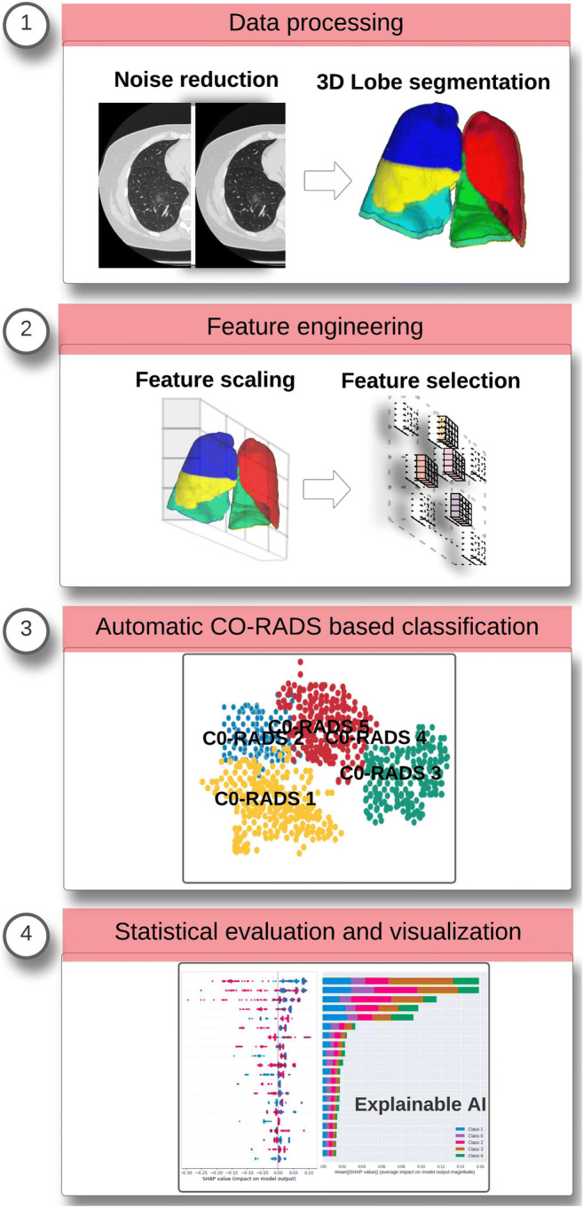
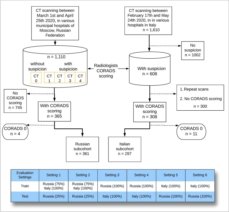
Methods: This multi-institutional review boards-approved retrospective study included 2720 chest CT scans (mean age, 58 years [range 18-100 years]) from Italian and Russian patients. Three board-certified radiologists from three countries assessed randomly selected subcohorts from each population and provided CO-RADS-based annotations. CT radiomic features were extracted from the selected subcohorts after preprocessing steps like lung lobe segmentation and automatic noise reduction. We compared three machine learning models, logistic regression (LR), multilayer perceptron (MLP), and random forest (RF) for the automated CO-RADS classification. Model evaluation was carried out in two scenarios, first, training on a mixed multi-demographic subcohort and testing on an independent hold-out dataset. In the second scenario, training was done on a single demography and externally validated on the other demography.
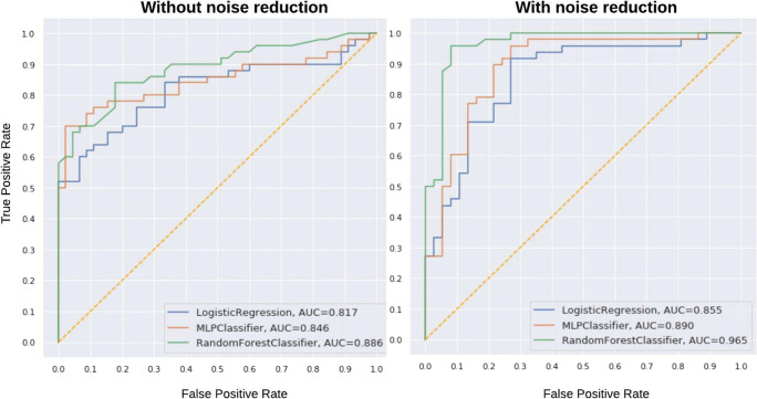
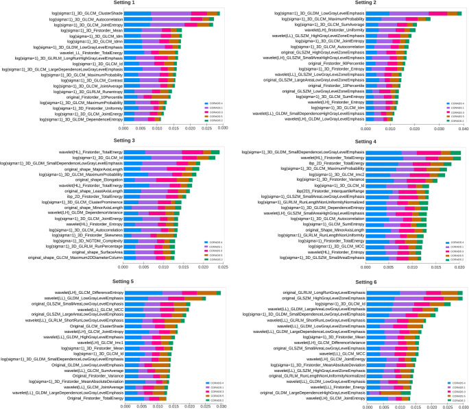
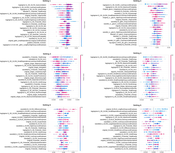
Results: The overall inter-observer agreement for the CO-RADS scoring between the radiologists was substantial (k = 0.80). Irrespective of the type of validation test scenario, suspected COVID-19 CT scans were identified with an accuracy of 84%. SHapley Additive exPlanations (SHAP) interpretation showed that the "wavelet_(LH)_GLCM_Imc1" feature had a positive impact on COVID prediction both with and without noise reduction. The application of noise reduction improved the overall performance between the classifiers for all types.
Conclusion: Using an automated model based on the COVID-19 Reporting and Data System (CO-RADS), we achieved clinically acceptable performance in a multi-demographic setting. This approach can serve as a standardized tool for automated COVID-19 assessment.
Keypoints: • Automatic CO-RADS scoring of large-scale multi-demographic chest CTs with mean AUC of 0.93 ± 0.04. • Validation procedure resembles TRIPOD 2b and 3 categories, enhancing the quality of experimental design to test the cross-dataset domain shift between institutions aiding clinical integration. • Identification of COVID-19 pneumonia in the presence of community-acquired pneumonia and other comorbidities with an AUC of 0.92.
Keywords: COVID-19; Deep learning; Diagnostic imaging; SARS-CoV-2; Tomography X-ray computed.
© 2022. The Author(s).
Conflict of interest statement
Matthijs Oudkerk holds a financial interest in the Institute of DiagNostic Accuracy Research (iDNA), an organization that aims to speed up the global implementation of the early detection of lung cancer with comorbidities in cardiovascular diseases and COPD.
The rest of the authors of this manuscript declare no relationships with any companies, whose products or services may be related to the subject matter of the article.
Figures
Similar articles
-
CT radiomics facilitates more accurate diagnosis of COVID-19 pneumonia: compared with CO-RADS.J Transl Med. 2021 Jan 7;19(1):29. doi: 10.1186/s12967-020-02692-3. J Transl Med. 2021. PMID: 33413480 Free PMC article.
-
Diagnostic accuracy and inter-observer agreement with the CO-RADS lexicon for CT chest reporting in COVID-19.Emerg Radiol. 2021 Dec;28(6):1045-1054. doi: 10.1007/s10140-021-01967-6. Epub 2021 Jul 24. Emerg Radiol. 2021. PMID: 34302561 Free PMC article.
-
Radiological Society of North America (RSNA) Expert Consensus Statement Related to Chest CT Findings in COVID-19 Versus CO-RADS: Comparison of Reporting System Performance Among Chest Radiologists and End-User Preference.Can Assoc Radiol J. 2021 Nov;72(4):806-813. doi: 10.1177/0846537120968919. Epub 2020 Nov 3. Can Assoc Radiol J. 2021. PMID: 33138634
-
Coronavirus disease 2019 (COVID-19) imaging reporting and data system (COVID-RADS) and common lexicon: a proposal based on the imaging data of 37 studies.Eur Radiol. 2020 Sep;30(9):4930-4942. doi: 10.1007/s00330-020-06863-0. Epub 2020 Apr 28. Eur Radiol. 2020. PMID: 32346790 Free PMC article.
-
GACDN: generative adversarial feature completion and diagnosis network for COVID-19.BMC Med Imaging. 2021 Oct 21;21(1):154. doi: 10.1186/s12880-021-00681-6. BMC Med Imaging. 2021. PMID: 34674660 Free PMC article. Review.
Cited by
-
Reproducibility of radiomics quality score: an intra- and inter-rater reliability study.Eur Radiol. 2024 Apr;34(4):2791-2804. doi: 10.1007/s00330-023-10217-x. Epub 2023 Sep 21. Eur Radiol. 2024. PMID: 37733025 Free PMC article. Clinical Trial.
-
Tracheal computed tomography radiomics model for prediction of the Omicron variant of severe acute respiratory syndrome coronavirus 2.Radiologie (Heidelb). 2024 Nov;64(Suppl 1):66-75. doi: 10.1007/s00117-024-01275-3. Epub 2024 Mar 6. Radiologie (Heidelb). 2024. PMID: 38446170 English.
-
Development of a machine learning model in prediction of the rapid progression of interstitial lung disease in patients with idiopathic inflammatory myopathy.Quant Imaging Med Surg. 2024 Dec 5;14(12):9258-9275. doi: 10.21037/qims-24-595. Epub 2024 Nov 8. Quant Imaging Med Surg. 2024. PMID: 39698644 Free PMC article.
References
-
- He Y. Translation: diagnosis and treatment protocol for novel coronavirus pneumonia (Trial Version 7) Infect Microbes Dis. 2020;2:48–54. doi: 10.1097/IM9.0000000000000022. - DOI
-
- Simpson S, Kay FU, Abbara S, et al. Radiological Society of North America expert consensus document on reporting chest CT findings related to COVID-19: endorsed by the Society of Thoracic Radiology, the American College of Radiology, and RSNA. Radiol Cardiothorac Imaging. 2020;2:e200152. doi: 10.1148/ryct.2020200152. - DOI - PMC - PubMed
Publication types
MeSH terms
Grants and funding
LinkOut - more resources
Full Text Sources
Medical
Research Materials
Miscellaneous
