. 2022 Jun;388(3):565-581.
doi: 10.1007/s00441-022-03613-0. Epub 2022 Apr 1.

Bone tissue engineering using 3D silk scaffolds and human dental pulp stromal cells epigenetic reprogrammed with the selective histone deacetylase inhibitor MI192

Affiliations

Bone tissue engineering using 3D silk scaffolds and human dental pulp stromal cells epigenetic reprogrammed with the selective histone deacetylase inhibitor MI192

Kenny Man et al. Cell Tissue Res. 2022 Jun.

Abstract

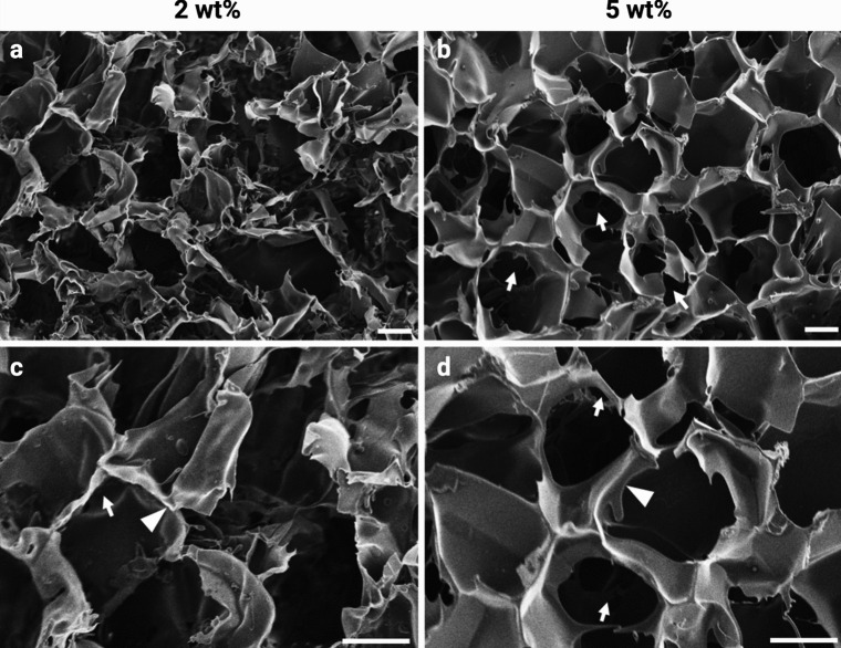
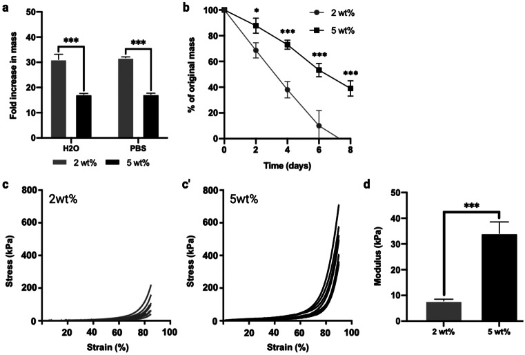
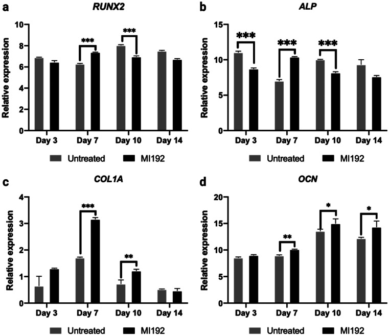
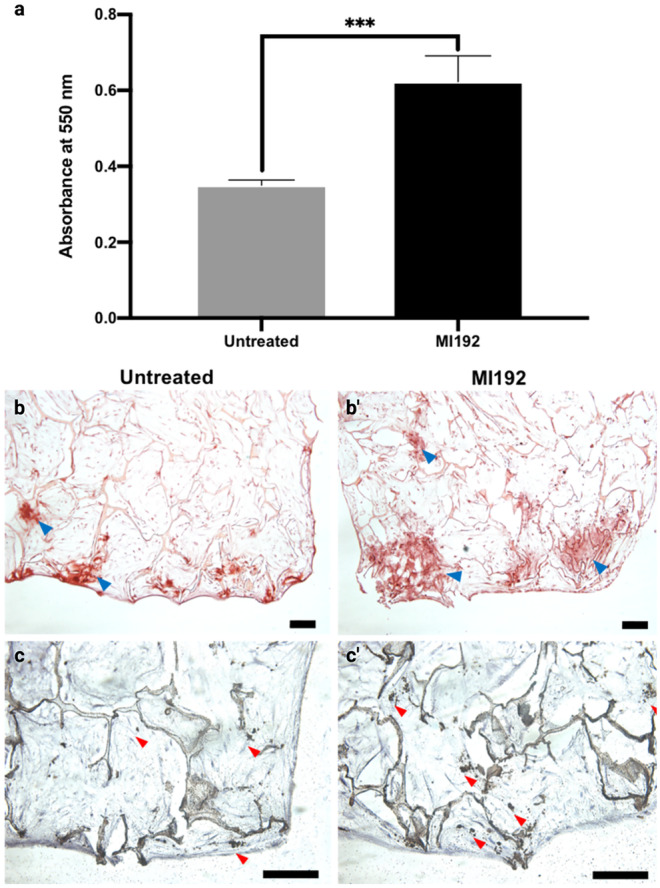
Epigenetics plays a critical role in regulating mesenchymal stem cells' (MSCs) fate for tissue repair and regeneration. There is increasing evidence that the inhibition of histone deacetylase (HDAC) isoform 3 can enhance MSC osteogenesis. This study investigated the potential of using a selective HDAC2 and 3 inhibitor, MI192, to promote human dental pulp stromal cells (hDPSCs) bone-like tissue formation in vitro and in vivo within porous Bombyx Mori silk scaffolds. Both 2 and 5 wt% silk scaffolds were fabricated and characterised. The 5 wt% scaffolds possess thicker internal lamellae, reduced scaffold swelling and degradation rates, whilst increased compressive modulus in comparison to the 2 wt% silk scaffold. MI192 pre-treatment of hDPSCs on 5 wt% silk scaffold significantly enhanced hDPSCs alkaline phosphatase activity (ALP). The expression of osteoblast-related genes (RUNX2, ALP, Col1a, OCN) was significantly upregulated in the MI192 pre-treated cells. Histological analysis confirmed that the MI192 pre-treated hDPSCs-silk scaffold constructs promoted bone extracellular matrix (ALP, Col1a, OCN) deposition and mineralisation compared to the untreated group. Following 6 weeks of subcutaneous implantation in nude mice, the MI192 pre-treated hDPSCs-silk scaffold constructs enhanced the vascularisation and extracellular matrix mineralisation compared to untreated control. In conclusion, these findings demonstrate the potential of using epigenetic reprogramming and silk scaffolds to promote hDPSCs bone formation efficacy, which provides evidence for clinical translation of this technology for bone augmentation.

Keywords: Bone tissue engineering; Epigenetics; Histone deacetylase inhibitor; Human dental pulp stromal cells; Silk scaffolds.


Conflict of interest statement

The authors declare no competing interests.

Figures

Fig. 1
Fig. 1
Experimental outline investigating the influence of MI192 pre-treatment on osteogenic differentiation of hDPSCs within 3D silk scaffold in vitro and in vivo. a The influence of altering silk concentration on the scaffolds physical properties and osteogenic capacity was evaluated. b The effect of MI192 in stimulating hDPSCs mineralisation within silk scaffolds in vitro was investigated. c Bone-like tissue formation induced by MI192 pre-treatment of hDPSCs was evaluated following 6 weeks of subcutaneous implantation in mice. (The figure was created with Biorender.com)
Fig. 2
Fig. 2
SEM micrographs showing the porosity, pore morphology and lamellae network in 2 wt% (a, c) and 5 wt% (b, d) silk scaffolds. The white arrows indicate the interconnection between the pores. The white arrowheads indicate the thin sheet-like lamellae between the pores. Scale bar, 100 μm
Fig. 3
Fig. 3
Effects of silk concentration on swelling capacity (a), in vitro degradation (b) and compressive modulus (c, c’, d). Data expressed as mean ± SD (n = 3). *P ≤ 0.05 and ***P ≤ 0.001
Fig. 4
Fig. 4
The influence of silk concentrations on hDPSC viability and osteogenic capacity. Live fluorescent staining of untreated/MI192 pre-treated hDPSCs after 24 h (aa’’’) and 6 weeks (bb’’’) in osteogenic culture. Low magnification (the top panel) scale bars = 200 μm, and high magnification (the second panel) scale bars = 50 μm. c ALPSA of MI192 pre-treated cells on silk scaffolds following 2 weeks in osteogenic culture. Data represented as mean ± SD (n = 3). ***P ≤ 0.001
Fig. 5
Fig. 5
The effects of MI192 pre-treatment on the expression of osteoblast-related genes (a-d) in hDPSCs cultured in 5 wt% silk scaffolds during osteogenic culture. a RUNX2, b ALP, c COL1A, d OCN. Gene expression was relative to the housekeeping gene GAPDH. Data represented as mean ± SD (n = 3). *P ≤ 0.05, **P ≤ 0.01 and ***P ≤ 0.001
Fig. 6
Fig. 6
Histological staining for picrosirius red and alcian blue (aa’’’) and immunohistochemical staining for ALP (bb’’’), Col1a (cc’’’) and OCN (dd’’’) of MI192 pre-treated/untreated hDPSCs on 5 wt% silk scaffolds (n = 3) after 6 weeks osteogenic culture in vitro. Low magnification scale bars = 200 μm (ad and a’’’ to d’’’), and high magnification scale bars = 50 μm (a’ to d’ and a’’ to d’’)
Fig. 7
Fig. 7
Effects of MI192 pre-treatment on hDPSCs calcium accumulation and mineralisation within 5 wt% silk scaffolds after 6 weeks osteogenic culture. a Quantitative alizarin red analysis, b & b’ alizarin red staining (red colour: blue arrowheads), c and c’) Von Kossa staining (black colour: red arrowheads). Data represented as mean ± SD (n = 3). ***P ≤ 0.001. Scale bar, 100 μm
Fig. 8
Fig. 8
Macroscopic and histological analysis for angiogenesis and scaffold degradation of MI192 pre-treated hDPSCs-silk constructs following 6 weeks subcutaneous implantation in mice. a and a’ Macroscopic image of cell-laden construct immediately prior to extraction. Light blue arrowheads highlight blood vessel formation. b and b’ H&E staining showing hDPSCs throughout the silk constructs with subcutaneous tissues at the periphery. Black arrowheads highlight blood vessel infiltration. Scale bars = 100 μm. c Blood vessel density in hDPSC-silk scaffolds quantified from histological images. d Internal lamellae thickness. Data represented as mean ± SD (n = 3). ** P ≤ 0.01
Fig. 9
Fig. 9
Histological and immunohistochemical staining of extracellular bone matrix and mineralisation of MI192 pre-treated hDPSCs within silk constructs following 6 weeks subcutaneous implantation in mice. Positive histological staining for collagen (red colour, picrosirius red/alcian blue staining) and black arrowheads indicated vessel formation (a, a’). Mineral nodule formation (black colour — light blue arrows, Von Kossa staining) (b, b’). Production of type I collagen (c, c’) and OCN (d, d’) proteins was evaluated via immunohistochemical staining (brown colour). Scale bar, 100 μm

References

    1. Abbott A. Cell culture: biology’s new dimension. Nature. 2003;424:870–872. doi: 10.1038/424870a. - DOI - PubMed
    1. Allmeling C, Jokuszies A, Reimers K, Kall S, Choi CY, Brandes G, Kasper C, Scheper T, Guggenheim M, Vogt PM. Spider silk fibres in artificial nerve constructs promote peripheral nerve regeneration. Cell Proliferat. 2008;41:408–420. doi: 10.1111/j.1365-2184.2008.00534.x. - DOI - PMC - PubMed
    1. Amini AR, Laurencin CT, Nukavarapu SP. Bone tissue engineering: recent advances and challenges. Crit Rev Biomed Eng. 2012;40:363–408. doi: 10.1615/CritRevBiomedEng.v40.i5.10. - DOI - PMC - PubMed
    1. Baker BM, Chen CS. Deconstructing the third dimension - how 3D culture microenvironments alter cellular cues. J Cell Sci. 2012;125:3015–3024. - PMC - PubMed
    1. Balasubramanian S, Verner E, Buggy JJ. Isoform-specific histone deacetylase inhibitors: the next step? Cancer Lett. 2009;280:211–221. doi: 10.1016/j.canlet.2009.02.013. - DOI - PubMed