. 2022 Apr;8(13):eabh1824.
doi: 10.1126/sciadv.abh1824. Epub 2022 Apr 1.

Deubiquitinase CYLD acts as a negative regulator of dopamine neuron survival in Parkinson's disease

Affiliations

Deubiquitinase CYLD acts as a negative regulator of dopamine neuron survival in Parkinson's disease

Sheila K Pirooznia et al. Sci Adv. 2022 Apr.

Abstract

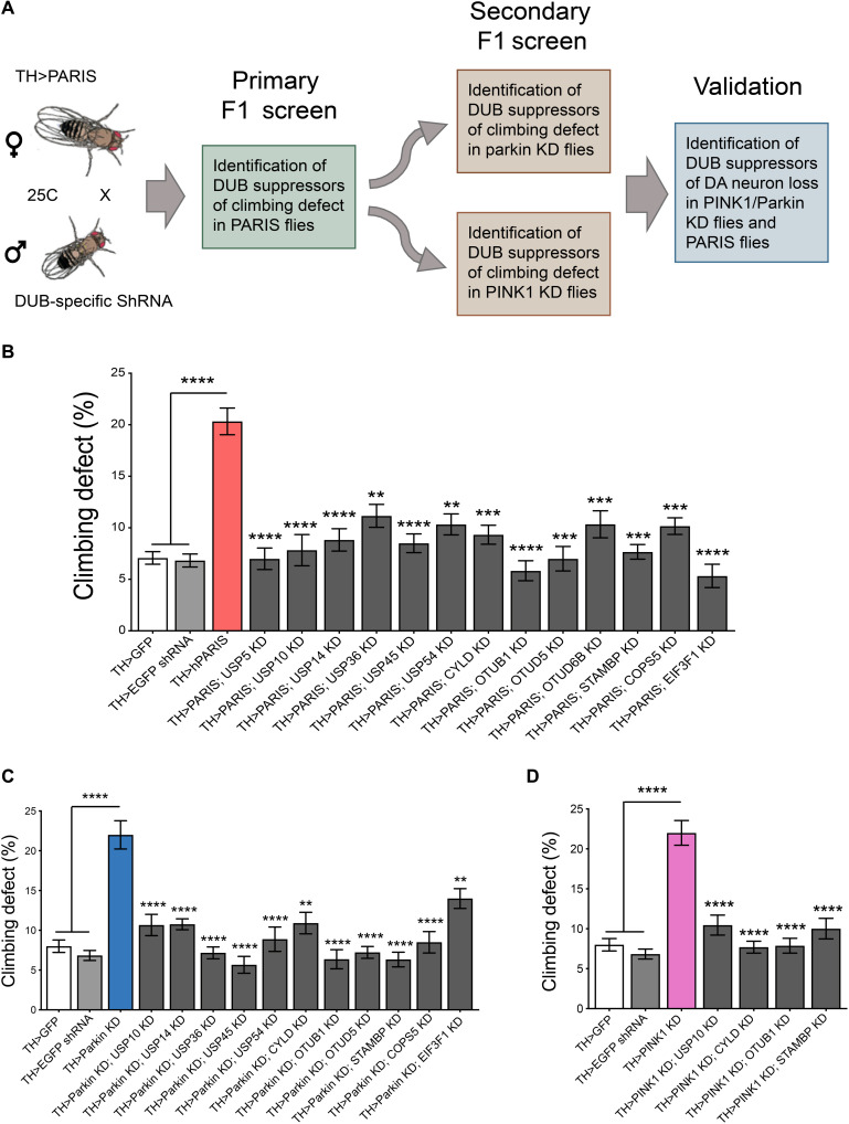
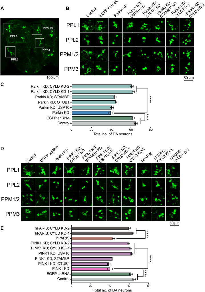
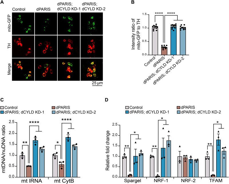
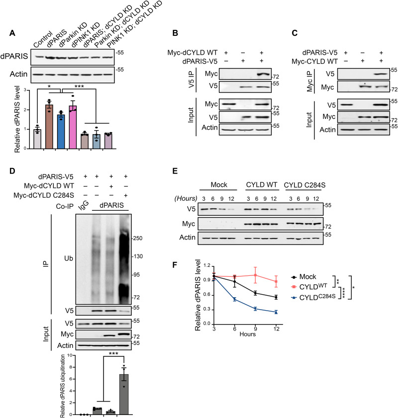
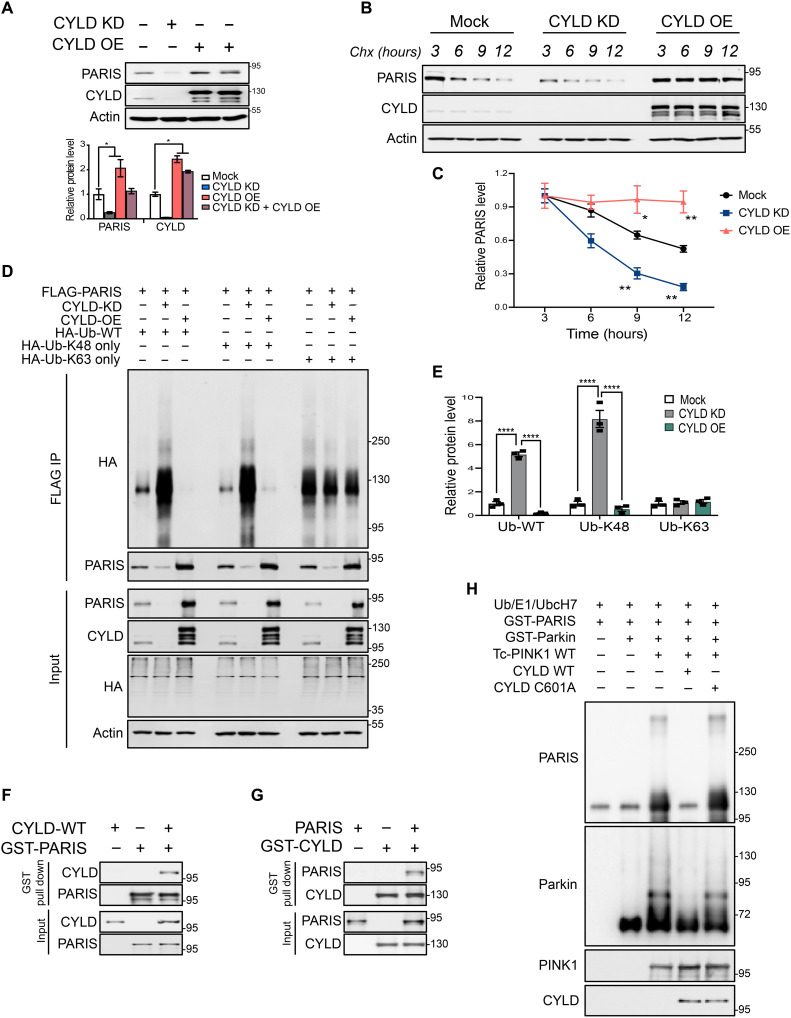
Mutations in PINK1 and parkin highlight the mitochondrial axis of Parkinson's disease (PD) pathogenesis. PINK1/parkin regulation of the transcriptional repressor PARIS bears direct relevance to dopamine neuron survival through augmentation of PGC-1α-dependent mitochondrial biogenesis. Notably, knockout of PARIS attenuates dopaminergic neurodegeneration in mouse models, indicating that interventions that prevent dopaminergic accumulation of PARIS could have therapeutic potential in PD. To this end, we have identified the deubiquitinase cylindromatosis (CYLD) to be a regulator of PARIS protein stability and proteasomal degradation via the PINK1/parkin pathway. Knockdown of CYLD in multiple models of PINK1 or parkin inactivation attenuates PARIS accumulation by modulating its ubiquitination levels and relieving its repressive effect on PGC-1α to promote mitochondrial biogenesis. Together, our studies identify CYLD as a negative regulator of dopamine neuron survival, and inhibition of CYLD may potentially be beneficial in PD by lowering PARIS levels and promoting mitochondrial biogenesis.


Figures

Fig. 1.
Fig. 1.. Genome-wide RNAi screen to identify candidate DUBs that function in PINK1/parkin pathway.
(A) Schematic of the screen. TH-Gal4–driven DUB-specific RNAi fly lines were used in an F1 primary screen to identify DUBs that rescued hPARIS-induced climbing defects on day 20. Such candidate DUBs were subjected to additional secondary F1 screens probing for DUBs that rescued climbing defects under conditions of DA parkin or PINK1 KD on day 20, respectively. Hits from the secondary screen were then examined for their ability to promote dopamine neuron survival in 20-day-old parkin or PINK1 KD flies. (B) Primary F1 screen based on rescue of PARIS-induced climbing defect identified 13 DUBs. (C) Summary of candidate DUBs from primary screen that rescued climbing defects in parkin KD flies. (D) Summary of candidate DUBs from primary screen that suppressed climbing defects in PINK1 KD flies and progressed for further validation. TH-Gal4/+ flies served as control. N = 60 flies per genotype for both primary and secondary screens. Quantitative data = means ± SEM. One-way analysis of variance (ANOVA); **P < 0.01, ***P < 0.001, ****P < 0.0001. See also figs. S1 and S2 and table S1.
Fig. 2.
Fig. 2.. Validation of candidate DUBs identified in primary and secondary RNAi screens places Drosophila cylindromatosis (dCYLD) in the PINK1/parkin pathway.
(A) Representative confocal image of the Drosophila whole brain showing the PPL1, PPL2, PPM1/2, and PPM3 DA neuron clusters. Scale bar, 100 μM. (B) Representative confocal images of individual dopamine neuron clusters visualized using TH immunofluorescence in the indicated genotypes under conditions of parkin KD. Scale bar, 50 μM. N = 10 flies per genotype. (C) Quantification of neuronal numbers within individual dopamine neuron clusters in the indicated genotypes. (D) Representative confocal images of individual dopamine neuron clusters visualized using TH immunofluorescence in the indicated genotypes under conditions of PINK1 KD or hPARIS overexpression. Scale bar, 50 μM. (E) Summary of dopamine neuron quantifications in the indicated genotypes. TH-Gal4/+ flies served as control. N = 10 flies per genotype. Quantitative data = means ± SEM. One-way ANOVA; ****P < 0.0001. See also fig. S3.
Fig. 3.
Fig. 3.. dCYLD KD promotes mitochondrial biogenesis in dopamine neurons.
(A) Representative confocal images showing mito-GFP (green)–labeled mitochondria within dopamine neurons immunostained for TH (red) in the indicated genotypes. Scale bar, 25 μM. (B) Quantification of intensity ratio of mito-GFP to TH immunofluorescence in the indicated genotypes. TH>mito-GFP flies served as control, N = 10 flies per genotype. (C) Assessment of mitochondrial DNA copy number in FACS-sorted dopamine neurons in 30-day-old flies of the indicated genotypes. Mean ratio of mtDNA to nuDNA from three independent FACS experiments, each using 50 whole fly brains shown. TH>GFP flies served as control. (D) Quantitative RT-PCR analysis of Drosophila homologs of PGC-1α (Spargel), NRF1 (ewg), NRF-2 (Delg), and TFAM in FACS-sorted DA neurons from 30-day-old flies. Mean values from three independent FACS experiments shown. TH>GFP flies served as control. Quantitative data = means ± SEM. One-way ANOVA; *P < 0.05, **P < 0.01, and ****P < 0.0001.
Fig. 4.
Fig. 4.. KD of dCYLD promotes dopamine neuron survival by destabilizing dPARIS through its DUB activity.
(A) Representative immunoblot and dPARIS quantification in flies expressing the indicated transgenes under the control of TH-Gal4 driver. TH-Gal4/+ flies served as control, N = 3. (B) Coimmunoprecipitation using anti-V5 antibodies shows interaction between C-terminal V5-tagged dPARIS and N-terminal Myc-tagged dCYLD in Drosophila S2 cells. Similar results were observed in three independent experiments. (C) Reciprocal coimmunoprecipitation experiments using anti-Myc antibodies verify dPARIS interaction with dCYLD in S2 cells transfected with indicated constructs in three independent experiments, N = 3. (D) Deubiquitination of dPARIS by dCYLD as assessed in S2 cells transfected with indicated constructs. Immunoblot analysis and relative quantification of immunoprecipitated (IP) dPARIS shows increased ubiquitination (Ub) of dPARIS in the presence of the dCYLD C284S catalytic mutant, thereby enhancing its proteasomal degradation, N = 3. (E) DUB activity of dCYLD affects the turnover rate of dPARIS. (F) Immunoblot analysis and dPARIS quantification in S2 cells at the indicated time points shows that while the dCYLD catalytic mutant accelerates dPARIS turnover, overexpression of WT dCYLD increases its half-life, N = 3. Quantitative data = means ± SEM. One-way ANOVA; *P < 0.05, **P < 0.01, ***P < 0.001, ****P < 0.0001. See also fig. S4.
Fig. 5.
Fig. 5.. Mammalian PARIS is a deubiquitination substrate of CYLD.
(A) Immunoblot analysis and quantification of endogenous PARIS in SH-SY5Y neuroblastoma cell line following CRISPR-Cas9–mediated KD or overexpression (OE) of CYLD, N = 3. (B) Cycloheximide chase experiment at the indicated time points showing PARIS turnover rate under conditions of CYLD KD or OE in SH-SY5Y cells. (C) Relative quantification of endogenous PARIS shows accelerated protein turnover under conditions of CYLD KD, whereas CYLD OE increases PARIS half-life. N = 3 independent experiments. (D) CYLD promotes deubiquitination of PARIS through hydrolysis of K48 ubiquitin chains. Ubiquitination of immunoprecipitated FLAG-tagged PARIS in SH-SY5Y cells monitored by immunoblot analysis under conditions of CYLD KD or OE in the presence of HA-tagged WT ubiquitin or ubiquitin mutants that can only append either K48 or K63 ubiquitin chains. (E) Quantification of PARIS ubiquitination showing enrichment of WT or K48 ubiquitin chains under conditions of CYLD KD but not CYLD OE. No changes in levels of K63 ubiquitin chains on PARIS observable under the different conditions assayed. N = 3. (F) GST pull-down assays using purified recombinant GST-tagged PARIS and WT CYLD indicate a robust interaction between PARIS and CYLD. Similar results were observed in three independent pull-down experiments. (G) Reciprocal GST pull-down assays using GST-tagged CYLD and PARIS confirm the direct interaction between PARIS and CYLD. N = 3. (H) In vitro deubiquitination of PARIS by CYLD. Immunoblot analysis of purified recombinant GST-PARIS ubiquitinated in the presence of PINK1 and activated parkin showing decreased ubiquitination in the presence of WT CYLD. The CYLD C601A catalytic mutant, however, has no impact on PARIS ubiquitination. Similar results were observed in three independent experiments. Quantitative data = means ± SEM. One-way ANOVA; *P < 0.05, **P < 0.01, and ****P < 0.0001. See also fig. S5.
Fig. 6.
Fig. 6.. CYLD KD prevents DA neurodegeneration in adult conditional parkin KD mice.
(A) Representative confocal images showing CYLD coexpression in TH-immunostained SN dopamine neurons in midbrain sections from WT C57BL/6 mice. Scale bar, 20 μM. (B) Schematic of experimental schedule following stereotaxic injection with AAV-GFP or AAV-GFP Cre in SN of 2-month-old C57BL6 mice of indicated genotype. (C) Representative immunofluorescence images verifying stereotaxic injection quality. Scale bar, 500 μM. (D) Representative immunoblots of indicated proteins in ventral midbrain lysates from adult mice harboring conditional KD of parkin, CYLD, or a combination of parkin and CYLD. (E) Relative quantification of PARIS, parkin, and CYLD in ventral midbrain lysates from floxed mice of indicated genotypes stereotaxically injected with AAV-GFP or AAV-GFP-Cre. Mean values from four independent stereotaxic injections in each case shown. (F) Relative quantification of PGC-1α and the indicated mitochondrial proteins in ventral midbrain lysates as shown in (C). N = 4 independent stereotaxic injections with AAV-GFP or AAV-GFP-Cre. (G) Representative TH immunostaining of midbrain sections from SN of mice homozygous for the floxed parkin, CYLD, or parkin; CYLD alleles injected with AAV-GFP or AAV-GFP-Cre. (H) Stereotaxic assessment of TH- and Nissl-positive neurons in the SN pars compacta (SNpc) of indicated injection groups (N = 8 unilaterally injected mice per group). Quantitative data = means ± SEM. One-way ANOVA with Tukey post hoc test; *P < 0.05, **P < 0.01, ***P < 0.001, and ****P < 0.0001. See also fig. S6.
Fig. 7.
Fig. 7.. CYLD down-regulation attenuates PARIS-mediated neurotoxicity in hES cell–derived midbrain dopamine neurons deficient for parkin activity.
(A) Immunoblot analysis of the indicated proteins in control (H1) and two independent parkin knockout (P-KO1 and P-KO2) human midbrain dopamine neurons. (B) Quantification of indicated proteins in the different conditions. Mean values from three independent experiments shown. (C) Quantification of indicated mitochondrial markers in the different conditions. N = 3 independent experiments. (D) Quantification of TH-positive dopamine neurons in differentiated human midbrain cultures immunostained with TH and neuronal TUJ1 marker. N = 6 independent experiments (E) Immunoblot analysis of mitochondrial biomarkers in indicated conditions. (F) Quantification of indicated proteins under respective conditions. N = 3 independent experiments. (G) Immunoblot analysis and quantification of LC3II/I ratio in differentiated human midbrain neurons. N = 3 independent experiments. (H) Representative confocal images of human midbrain dopamine neurons immunostained for TH (green) and puromycin. Puromycin-labeled mitochondrial proteins detected by colocalization of anti-puromycin immunofluorescence (magenta) with the mitochondrial marker Tomm20 (red). (I) Quantification of anti-puromycin colocalization with Tomm20 in TH-positive midbrain dopamine neurons. Mean values from at least 15 TH-positive midbrain dopamine neurons from three independent differentiation shown. (J) Representative confocal images of human midbrain dopamine neurons expressing SNAP-Tag-Cox8a fusion protein immunostained for TH (magenta). Existing mitochondrial pool (old mito) shown in red and mitochondria newly assembled (new mito) shown in green. Nucleus stained with 4′,6-diamidino-2-phenylindole (DAPI) (blue). (K) Ratio of newly assembled to old mitochondria in TH-positive midbrain dopamine neurons. Mean values from at least 15 TH-positive midbrain dopamine neurons from three independent differentiations shown. (L) Immunoblot analysis and quantification of CYLD expression in SN from postmortem PD and age-matched controls. N = 4 per group. Quantitative data = means ± SEM. One-way ANOVA; *P < 0.05, **P < 0.01, ***P < 0.001, and ****P < 0.0001.

Similar articles

Cited by

References

    1. Kitada T., Asakawa S., Hattori N., Matsumine H., Yamamura Y., Minoshima S., Yokochi M., Mizuno Y., Shimizu N., Mutations in the parkin gene cause autosomal recessive juvenile parkinsonism. Nature 392, 605–608 (1998). - PubMed
    1. Valente E. M., Abou-Sleiman P. M., Caputo V., Muqit M. M., Harvey K., Gispert S., Ali Z., Del Turco D., Bentivoglio A. R., Healy D. G., Albanese A., Nussbaum R., Gonzalez-Maldonado R., Deller T., Salvi S., Cortelli P., Gilks W. P., Latchman D. S., Harvey R. J., Dallapiccola B., Auburger G., Wood N. W., Hereditary early-onset Parkinson’s disease caused by mutations in PINK1. Science 304, 1158–1160 (2004). - PubMed
    1. Kane L. A., Lazarou M., Fogel A. I., Li Y., Yamano K., Sarraf S. A., Banerjee S., Youle R. J., PINK1 phosphorylates ubiquitin to activate Parkin E3 ubiquitin ligase activity. J. Cell Biol. 205, 143–153 (2014). - PMC - PubMed
    1. Koyano F., Okatsu K., Kosako H., Tamura Y., Go E., Kimura M., Kimura Y., Tsuchiya H., Yoshihara H., Hirokawa T., Endo T., Fon E. A., Trempe J. F., Saeki Y., Tanaka K., Matsuda N., Ubiquitin is phosphorylated by PINK1 to activate parkin. Nature 510, 162–166 (2014). - PubMed
    1. Scarffe L. A., Stevens D. A., Dawson V. L., Dawson T. M., Parkin and PINK1: Much more than mitophagy. Trends Neurosci. 37, 315–324 (2014). - PMC - PubMed

MeSH terms