Review
. 2022 Jul 8:45:339-360.
doi: 10.1146/annurev-neuro-111020-105359. Epub 2022 Apr 1.

Neuroimmune Interactions in Peripheral Organs

Affiliations
Review

Neuroimmune Interactions in Peripheral Organs

Roel G J Klein Wolterink et al. Annu Rev Neurosci. .

Abstract

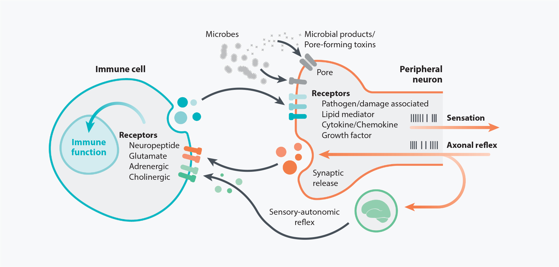
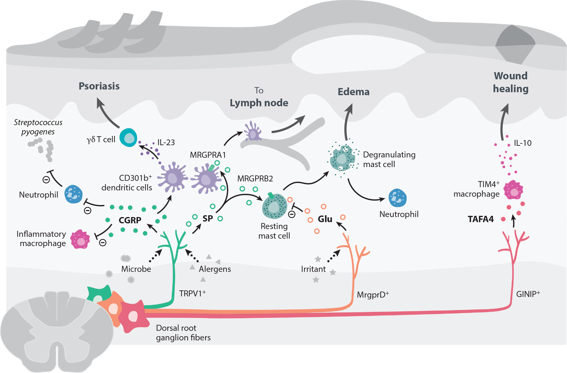
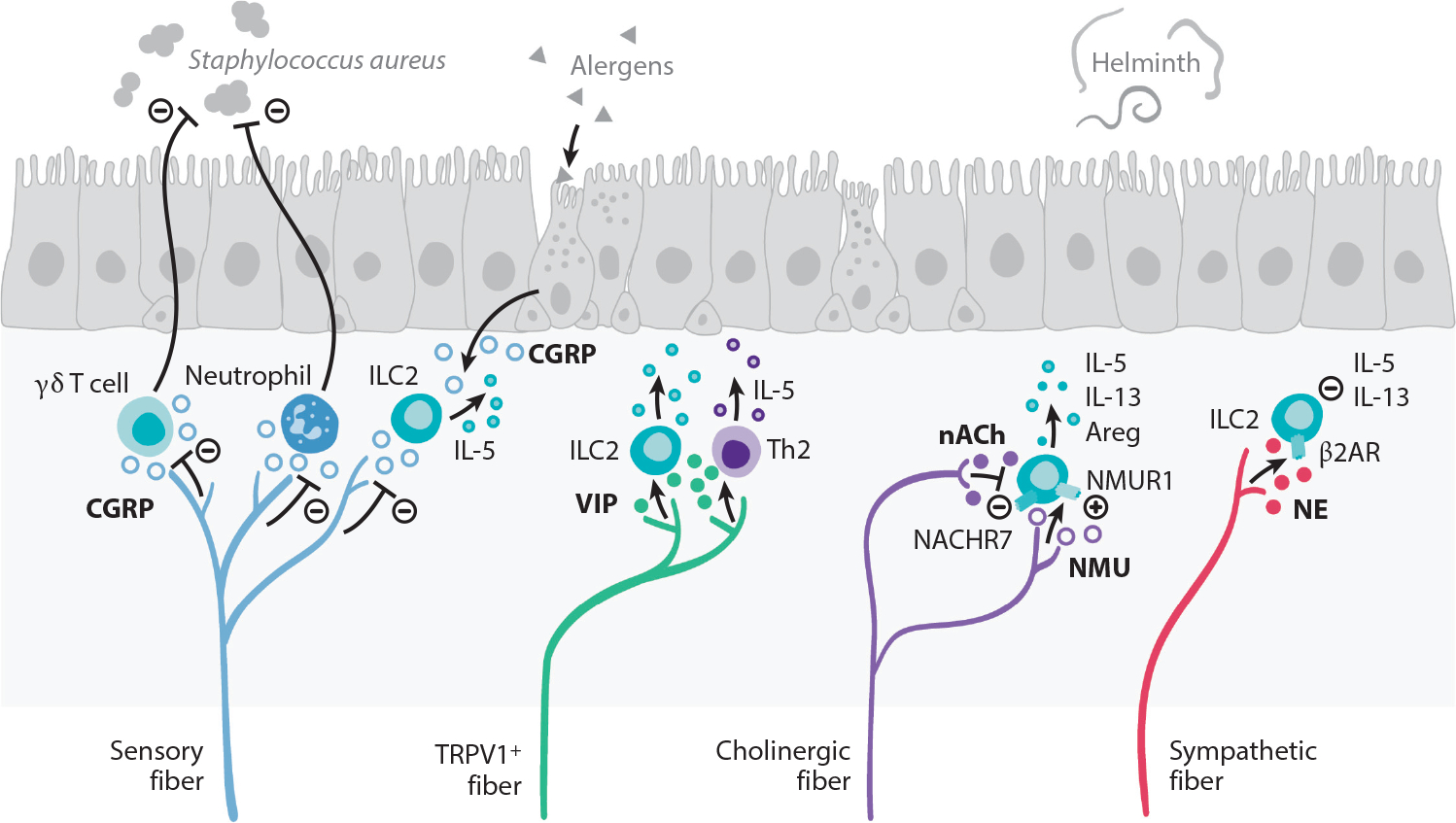
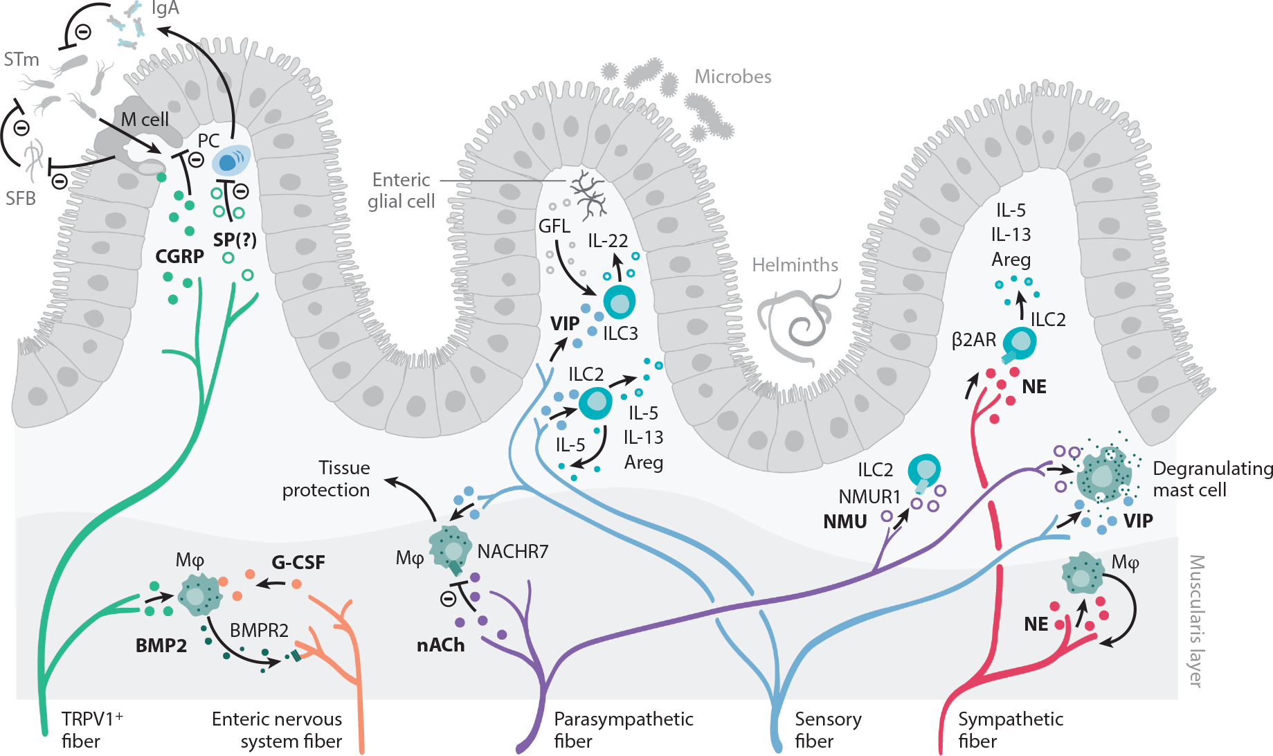
Interactions between the nervous and immune systems were recognized long ago, but recent studies show that this crosstalk occurs more frequently than was previously appreciated. Moreover, technological advances have enabled the identification of the molecular mediators and receptors that enable the interaction between these two complex systems and provide new insights on the role of neuroimmune crosstalk in organismal physiology. Most neuroimmune interactions occur at discrete anatomical locations in which neurons and immune cells colocalize. Here, we describe the interactions of the different branches of the peripheral nervous system with immune cells in various organs, including the skin, intestine, lung, and adipose tissue. We highlight how neuroimmune crosstalk orchestrates physiological processes such as host defense, tissue repair, metabolism, and thermogenesis. Unraveling these intricate relationships is invaluable to explore the therapeutic potential of neuroimmune interactions.

Keywords: mucosal immunology; neuroimmune interactions; neuroimmunology.


Conflict of interest statement

DISCLOSURE STATEMENT

I.M.C. is on scientific advisory boards for GSK Pharmaceuticals and LiMM Therapeutics. His lab receives funding from Allergan Pharmaceuticals for sponsored research. H.V.-F. is on the board of LiMM Therapeutics.

Figures

Figure 1
Figure 1
Direct communication between peripheral neurons and immune cells. Immune cell function can be modulated by factors produced from the PNS, as many immune cell types express receptors for neuropeptides and neurotransmitters. Reciprocally, the PNS can be regulated by the immune system since peripheral nerve terminals express canonical immune-related receptors for molecules such as cytokines, chemokines, and lipids. Nociceptive neurons also express receptors that detect microbes and/or their products, thereby allowing the nervous system to sense potential infections. In response to environmental cues such as inflammatory cytokines acting on receptors expressed by peripheral nerves, action potentials are generated to produce a variety of neuronal outputs, including activating reflex circuits and sensations such as pain and itch. One potential outcome is an antidromic axonal reflex that results in synaptic vesicle fusion and release of neuronal factors, which can in turn affect immune cell behavior and function. Signals from peripheral sensory nerves can also integrate with the central nervous system to produce a sensory-autonomic reflex and synaptic vesicle release. Abbreviation: PNS, peripheral nervous system.
Figure 2
Figure 2
Peripheral neuroimmune interactions in the skin. Most of the skin is innervated by sensory fibers originating from dorsal root ganglia, which have been shown to communicate with skin-resident and infiltrating immune cells. In bacterial infections caused by Staphylococcus aureus or Streptococcus pyogenes, pore-forming toxins activate Nav1.8+ and TRPV1+ nociceptor neurons to release CGRP. CGRP can subsequently suppress macrophages from producing inflammatory cytokines like TNFα and decrease neutrophil recruitment and function. This inhibitory activity of CGRP leads to the loss of control of bacterial infection. TRPV1+ fibers can also be activated by Candida albicans to release CGRP during infection. CGRP acts on dermal CD301b+ DCs to produce IL-23, which promotes dermal γδ T cell proliferation and secretion of IL-17 to control fungal infection. However, this DC/γδ T cell/IL-17 circuit can also promote inflammation and pathology in a mouse model of psoriasis. Active proteases in allergens trigger TRPV1+ neurons to release SP, which acts on MRGPRA1 on DCs to promote their migration to draining lymph nodes and prime a Th2 immune response. SP release from TRPV1+ sensory neurons also trigger mast cell degranulation via binding MRGPRB2. The SP-MRGPRB2 mast cell axis drives tissue edema and neutrophil recruitment. By contrast, nonpeptidergic MRGPRD+ sensory neurons release glutamate to suppress mast cell degranulation. GINIP+ sensory fibers promote tissue healing by releasing the neuropeptide TAFA4 to promote release of the anti-inflammatory cytokine IL-10 by TIM4+ macrophages. Abbreviations: CGRP, calcitonin gene–related peptide; DC, dendritic cell; GINIP, Gαi-interacting protein; MRGPR, Mas-related G protein–coupled receptor; Nav, voltage-gated sodium channel; SP, substance P; TAFA4, TAFA chemokine like family member 4; Th2, T-helper type 2; TIM4, T cell immunoglobulin and mucin domain containing 4; TNFα, tumor necrosis factor alpha; TRPV1, transient receptor potential cation channel V member 1.
Figure 3
Figure 3
Peripheral neuroimmune interactions in the lungs. The vagus nerve supplies most of the sensory and cholinergic innervation of the airways. Allergic inflammation in the lungs stimulates the production of the type 2 cytokine IL-5 that directly stimulates sensory neurons to release VIP. VIP recruits and activates ILC2s and CD4+ T cells, which further potentiate airway inflammation. TRPV1+ pulmonary fibers restrict antibacterial immunity through secretion of CGRP, which restricts recruitment of neutrophils and γδ T cells. CGRP, released by sensory fibers and neuroendocrine cells that are found interspersed in the lung epithelium, also regulates IL-5 production by ILC2s. Cholinergic fibers release the neuropeptide NMU, which is a potent activator of ILC2s via NMUR1 that responds by producing the type 2 effector cytokines IL-5, IL-13, and Areg that are important for helminth immunity. In turn, acetylcholine inhibits ILC2 function via NACHR7. Sympathetic fibers release NE that acts on β2AR expressed by ILC2s, downmodulating the production of IL-5 and IL-13. Abbreviations: β2AR, β2 adrenergic receptor; Areg, amphiregulin; CGRP, calcitonin gene–related peptide; ILC2, group 2 innate lymphoid cell; NACHR7, nicotinic acetylcholine receptor 7; NE, norepinephrine; NMU, neuromedin U; NMUR1, neuromedin U receptor 1; TRPV1, transient receptor potential cation channel V member 1; VIP, vasoactive intestinal peptide.
Figure 4
Figure 4
Peripheral neuroimmune interactions in the gut. The intestine is innervated by sensory fibers and the autonomic nervous system, composed of enteric neurons and sympathetic and parasympathetic fibers. Sensory afferents from vagal ganglia and lumbar-sacral DRG innervate the gastrointestinal tract and mediate protection to STm. M cells in Peyer’s patches and mucosa-associated lymphoid tissue facilitate STm transfer to establish an infection. Sensing of STm by DRG TRPV1+ and Nav1.8+ fibers cause CGRP release. CGRP acts on M cells to decrease their numbers and to limit STm uptake. M cells also negatively regulate SFB density, which further antagonizes STm infection. SP may also negatively regulate immune responses to STm, as NK1R-deficient mice exhibit enhanced levels of serum IgA and increased numbers of IgA-producing cells in the lamina propria. Sensory fibers also release VIP, which acts on ILC2s and ILC3s that release type 2 cytokines and tissue-protective IL-22, respectively. ILC3s are also stimulated by the RET ligand GFL, released by enteric glial cells. Enteric nervous system fibers release G-CSF, which promotes muscularis macrophage maintenance. In turn, macrophages produce BMP2, which activates neurons via the BMPR2. Furthermore, acetylcholine acts as a negative regulator of tissue-protective muscularis macrophages via the NACHR7. NE released by sympathetic fibers promotes macrophage-induced protection of neurons during infection. The function of intestinal ILC2 is also regulated by NMU, which acts as a strong stimulator of ILC2 function, and NE, which signals through the β2AR. Finally, intestinal mast cells can integrate a wide variety of neuronal cues and respond by producing a range of inflammatory mediators, which is covered in other excellent reviews and is beyond the scope of this piece. Abbreviations: β2AR, β2 adrenergic receptor; Areg, amphiregulin; BMP2, bone morphogenetic protein 2; BMPR2, bone morphogenetic protein receptor type 2; CGRP, calcitonin gene–related peptide; DRG, dorsal root ganglia; G-CSF, granulocyte colony stimulating factor; GFL, GDNF family of ligands; IgA, immunoglobulin A; ILC2, group 2 innate lymphoid cell; ILC3, group 3 innate lymphoid cell; M, microfold; Mϕ, macrophage; NACHR7, nicotinic acetylcholine receptor 7; Nav, voltage-gated sodium channel; NE, norepinephrine; NK1R, neurokinin-1 receptor; NMU, neuromedin U; NMUR1, neuromedin U receptor; PC, Plasma cell; RET, rearranged during transfection; SFB, segmented filamentous bacteria; SP, substance P; STm, Salmonella enterica serovar Typhimurium; TRPV1, transient receptor potential cation channel subfamily V member 1; VIP, vasoactive intestinal peptide.
Figure 5
Figure 5
Peripheral neuroimmune interactions in the adipose tissue. (a) SAMs localized in adipose tissue around sympathetic fibers express the NE transporter SLC6A2 and the degradation enzyme MAOA. Thus, these macrophages act to remove NE from the interstitium, thereby lowering the amount of NE available to adipocytes. Normally, NE stimulates adipocyte lipolysis and fatty acid oxidation, giving rise to beige adipocytes in a process called browning. (b) Aging stimulates the NLRP3 inflammasome, inducing GDF3 that promotes MAOA expression in NE-degrading macrophages. Aging dampens NE release and lipolysis in adipose tissue through increased removal of NE, contributing to increased availability of fatty acids, which are required to survive starvation and to tolerate exercise. (c) A sympathetic aorticorenal-adipose circuit connects to the brain and regulates group 2 innate lymphoid cells (ILC2s) via mesenchymal stem cells. Gonadal adipose tissue mesenchyme units translate NE cues to expression of the RET ligand GDNF. In turn, this neurotrophic fact controls adipose tissue ILC2 function via the neuroregulatory receptor RET. ILC2s control adipocyte metabolism via the effector cytokines IL-5, IL-13, and Met-Enk. Abbreviations: β2AR, β2 adrenergic receptor; ARG, aorticorenal ganglion; Enk, enkephalin; GDF3, growth differentiation factor 3; GDNF, glial-derived neurotrophic factor; ILC2, group 2 innate lymphoid cell; MAOA, monoamine oxidase A; MSC, mesenchymal stem cell; NE, norepinephrine; NLRP3, NLR family pyrin domain containing 3; PVH, paraventricular nucleus of hypothalamus; RET, rearranged during transfection; SAM, sympathetic neuron–associated macrophage.

References

    1. Afan AM, Broome CS, Nicholls SE, Whetton AD, Miyan JA. 1997. Bone marrow innervation regulates cellular retention in the murine haemopoietic system. Br. J. Haematol. 98(3):569–77 - PubMed
    1. Andersson RG, Grundström N. 1987. Innervation of airway smooth muscle. Efferent mechanisms. Pharmacol. Ther. 32(2):107–30 - PubMed
    1. Arranz L, Sánchez-Aguilera A, Martín-Pérez D, Isern J, Langa X, et al. 2014. Neuropathy of haematopoietic stem cell niche is essential for myeloproliferative neoplasms. Nature 512(7512):78–81 - PubMed
    1. Bai A, Lu N, Guo Y, Chen J, Liu Z. 2009. Modulation of inflammatory response via α2-adrenoceptor blockade in acute murine colitis. Clin. Exp. Immunol. 156(2):353–62 - PMC - PubMed
    1. Baral P, Umans BD, Li L, Wallrapp A, Bist M, et al. 2018. Nociceptor sensory neurons suppress neutrophil and γδ T cell responses in bacterial lung infections and lethal pneumonia. Nat. Med. 24(4):417–26 - PMC - PubMed

Publication types

MeSH terms