. 2022 Apr 19;119(16):e2119467119.
doi: 10.1073/pnas.2119467119. Epub 2022 Apr 1.

Structural conservation among variants of the SARS-CoV-2 spike postfusion bundle

Affiliations

Structural conservation among variants of the SARS-CoV-2 spike postfusion bundle

Kailu Yang et al. Proc Natl Acad Sci U S A. .

Abstract

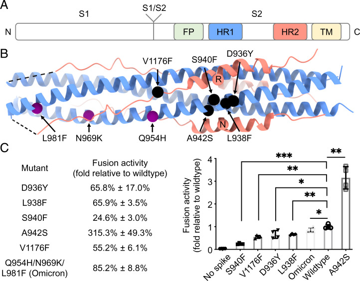
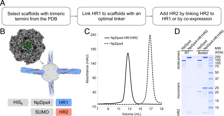
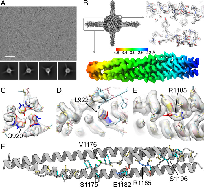
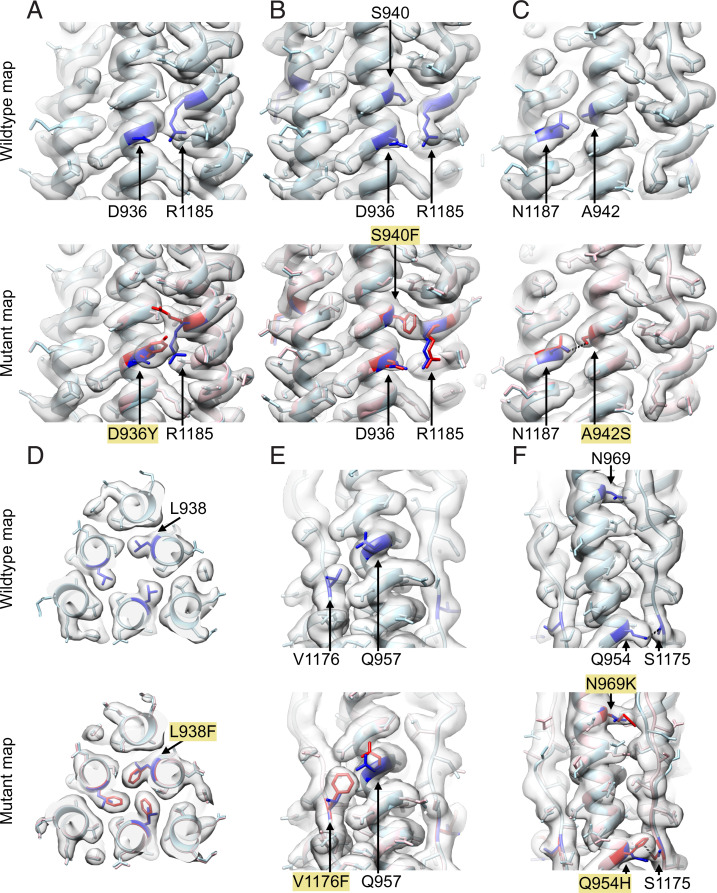
Variants of severe acute respiratory syndrome coronavirus 2 (SARS-CoV-2) challenge currently available COVID-19 vaccines and monoclonal antibody therapies due to structural and dynamic changes of the viral spike glycoprotein (S). The heptad repeat 1 (HR1) and heptad repeat 2 (HR2) domains of S drive virus–host membrane fusion by assembly into a six-helix bundle, resulting in delivery of viral RNA into the host cell. We surveyed mutations of currently reported SARS-CoV-2 variants and selected eight mutations, including Q954H, N969K, and L981F from the Omicron variant, in the postfusion HR1HR2 bundle for functional and structural studies. We designed a molecular scaffold to determine cryogenic electron microscopy (cryo-EM) structures of HR1HR2 at 2.2–3.8 Å resolution by linking the trimeric N termini of four HR1 fragments to four trimeric C termini of the Dps4 dodecamer from Nostoc punctiforme. This molecular scaffold enables efficient sample preparation and structure determination of the HR1HR2 bundle and its mutants by single-particle cryo-EM. Our structure of the wild-type HR1HR2 bundle resolves uncertainties in previously determined structures. The mutant structures reveal side-chain positions of the mutations and their primarily local effects on the interactions between HR1 and HR2. These mutations do not alter the global architecture of the postfusion HR1HR2 bundle, suggesting that the interfaces between HR1 and HR2 are good targets for developing antiviral inhibitors that should be efficacious against all known variants of SARS-CoV-2 to date. We also note that this work paves the way for similar studies in more distantly related viruses.

Keywords: COVID-19; HR1HR2; SARS-CoV-2; cryogenic electron microscopy; membrane fusion.


Conflict of interest statement

The authors declare no competing interest.

Figures

Fig. 1.
Fig. 1.
Mutations of interest in the HR1HR2 bundle of SARS-CoV-2 variants. (A) Schematic diagram of the domain structures of the SARS-CoV-2 spike protein. The N and C termini are labeled on the left and right, respectively. FP, fusion peptide; HR1, heptad repeat 1; HR2, heptad repeat 2; TM, transmembrane region. (B) Locations of the five selected point mutations of SARS-CoV-2 variants (black spheres) and the three mutations of the SARS-CoV-2 Omicron variant (purple spheres) indicated in the crystal structure of the HR1HR2 bundle (PDB ID code 6lxt). Two HR2 residues, R1185 and N1187, that may be affected by the selected mutations are shown as red spheres. The HR1 and HR2 fragments are colored as light blue and light red, respectively. (C) Effects on fusion activity of these mutations. The fusion activity is shown as a percentage (Left)/fold change (Right) relative to that of the wild type (Materials and Methods). The Omicron construct used here for the fusion assay has three mutations—Q954H, N969K, and L981F—in the HR1 portion of the HR1HR2 bundle, but not other mutations from different regions of the spike found in the Omicron variant. *P < 0.05, **P < 0.01, ***P < 0.001, by a Student’s t test.
Fig. 2.
Fig. 2.
Construct design and optimization of the scaffolded HR1HR2 bundle. (A) Workflow of designing and optimizing the scaffolded HR1HR2 bundle. (B) Diagram of construct design. (Top Left) Crystal structure of the dodecamer Dps4 from N. punctiforme (NpDps4, PDB ID code 5hjf), with one of its four three-helix-bundle termini shown as a green cartoon and the rest as a gray surface. (Middle) Cartoon of the scaffolded HR1HR2 bundle. (Bottom) Diagram of the constructs for coexpression of the scaffolded HR1HR2 bundle. (C) Size exclusion chromatography profiles of the purified scaffolded HR1HR2 bundle and the NpDps4 scaffold alone. (D) SDS-PAGE gel of the purified scaffolded HR1HR2 bundle and the scaffold alone, with or without boiling. The bands above the 100 kDa marker correspond to the respective dodecamers, the bands of corresponding monomers are around the 20 kDa and 25 kDa markers, and the band for the HR2 fragment alone is below the 10 kDa marker.
Fig. 3.
Fig. 3.
Cryo-EM studies of the scaffolded HR1HR2 complex. (A) Representative raw EM micrograph of the scaffolded wild-type HR1HR2 bundle and reference-free 2D class averages using a total of 2,896,745 selected particles (2D classification in cryoSPARC; SI Appendix, Fig. S4). The box size of the 2D class averages is 326.5 Å. (Scale bar on the raw micrograph, 500 Å.) (B) The globally refined map of the scaffolded HR1HR2 bundle shows the clearly resolved dodecameric NpDps4 scaffold region and four HR1HR2 complexes that are projecting from the scaffold with progressively increasing disorder. Symmetry expansion, signal subtraction, and local refinement resolve the HR1HR2 complexes, typically achieving resolutions ranging from 2.2–3.8 Å (SI Appendix, Fig. S4). (CE) Comparison of the EM map (gray) of the wild-type SARS-CoV-2 HR1HR2 complex (yellow, this study) with two published crystal structures of the HR1HR2 bundle (PDB ID codes 6xlt, pink, and 6m1v, light blue), focusing on the residues with discrepant side-chain conformations between the two crystal structures. Residues Q920, L922, and R1185 of the two crystal structures are colored as red and blue, respectively, to emphasize the differences. (F) Analysis of key interactions between the HR1 and HR2 fragments. Residues of HR2 that are involved in hydrophobic interactions with HR1 are colored yellow. Note that the variant mutation V1176 (colored green) is not interacting with HR1. Hydrogen bonds are shown as cyan dashed lines. Salt bridges are indicated by positively charged residues (colored blue) and negatively charged residues (colored red). Only one HR2 protomer and its neighboring two HR1 protomers are shown for clarity.
Fig. 4.
Fig. 4.
Local variation and global conservation of the structures of the studied mutants. (AF) The EM structures of the HR1HR2 D936Y, S940F, A942S, L938F, and V1176F single mutants and the Omicron HR1HR2 triple mutant are shown in A through F, respectively. In each panel, the wild-type structure (light blue) and wild-type map are shown on the top, and the wild-type and mutant (pink) structures, and the mutant map are shown on the bottom. The mutated residues and affected residues are colored blue for the wild-type structure and red for the mutant structures. The hydrogen bonds between S942 and N1187 (C), Q954 and S1175 (F), and H954 and S1175 (F) are shown as black dashed lines.

Similar articles

Cited by

References

    1. Zhou P., et al. , A pneumonia outbreak associated with a new coronavirus of probable bat origin. Nature 579, 270–273 (2020). - PMC - PubMed
    1. Manzanares-Meza L. D., Medina-Contreras O., SARS-CoV-2 and influenza: A comparative overview and treatment implications. Bol. Méd. Hosp. Infant. México 77, 262–273 (2020). - PubMed
    1. Wang P., et al. , Antibody resistance of SARS-CoV-2 variants B.1.351 and B.1.1.7. Nature 593, 130–135 (2021). - PubMed
    1. Wibmer C. K., et al. , SARS-CoV-2 501Y.V2 escapes neutralization by South African COVID-19 donor plasma. Nat. Med. 27, 622–625 (2021). - PubMed
    1. Abdool Karim S. S., de Oliveira T., New SARS-CoV-2 variants — Clinical, public health, and vaccine implications. N. Engl. J. Med. 384, 1866–1868 (2021). - PMC - PubMed

Substances

Supplementary concepts