Review
. 2022 Apr 21;82(8):1501-1513.
doi: 10.1016/j.molcel.2022.03.012. Epub 2022 Mar 31.

Mechanisms underlying ubiquitin-driven selective mitochondrial and bacterial autophagy

Affiliations
Review

Mechanisms underlying ubiquitin-driven selective mitochondrial and bacterial autophagy

Ellen A Goodall et al. Mol Cell. .

Abstract

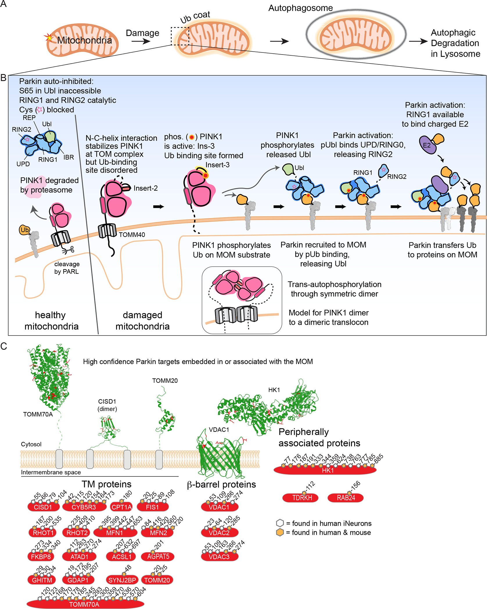
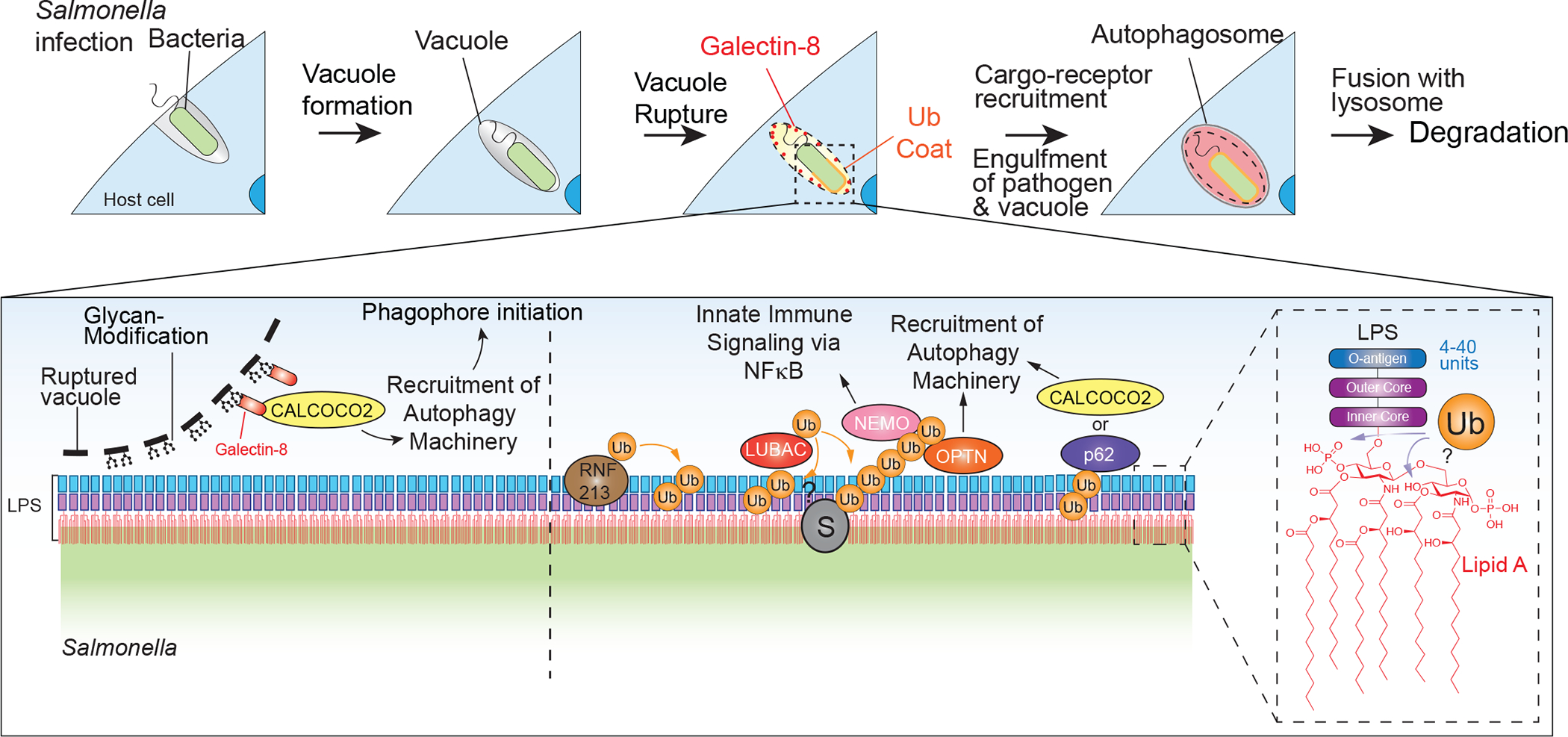
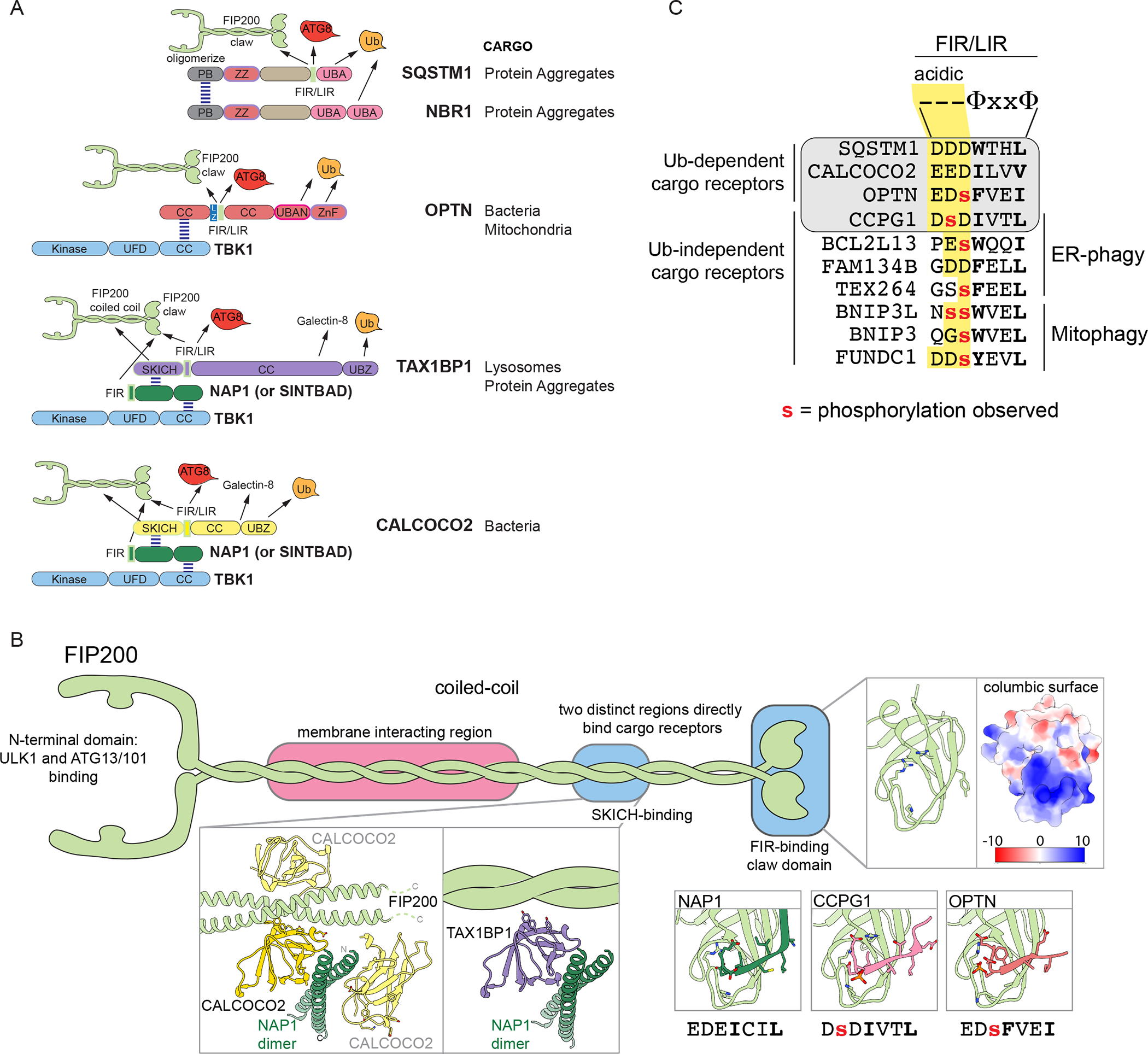
Selective autophagy specifically eliminates damaged or superfluous organelles, maintaining cellular health. In this process, a double membrane structure termed an autophagosome captures target organelles or proteins and delivers this cargo to the lysosome for degradation. The attachment of the small protein ubiquitin to cargo has emerged as a common mechanism for initiating organelle or protein capture by the autophagy machinery. In this process, a suite of ubiquitin-binding cargo receptors function to initiate autophagosome assembly in situ on the target cargo, thereby providing selectivity in cargo capture. Here, we review recent efforts to understand the biochemical mechanisms and principles by which cargo are marked with ubiquitin and how ubiquitin-binding cargo receptors use conserved structural modules to recruit the autophagosome initiation machinery, with a particular focus on mitochondria and intracellular bacteria as cargo. These emerging mechanisms provide answers to long-standing questions in the field concerning how selectivity in cargo degradation is achieved.

Keywords: cargo receptor; mitophagy; selective autophagy; ubiquitin; xenophagy.


Conflict of interest statement

Declaration of interests J.W.H. is a consultant and founder of Caraway Therapeutics and is a founding board member of Interline Therapeutics.

Figures

Figure 1.
Figure 1.. Regulated cargo ubiquitylation to promotes autophagic clearance of damaged mitochondria and intracellular bacteria.
(A) Schematic of Ub-dependent mitophagy. Damaged mitochondria elicits a signal resulting in creation of a Ub “coat” surrounding the mitochondria. This Ub coat functions to promote formation of an autophagosome, which then can fuse with the lysosome to promote mitochondrial clearance. (B) A feed-forward mechanism for Parkin and PINK1-dependent mitophagy. In cells with healthy mitochondria, cytosolic Parkin is in an auto-inhibited form and PINK1 is rapidly degraded. In response to mitochondrial damage, PINK1 is stabilized on the MOM in association with the translocon and trans-phosphorylates to generate a binding site for both Ub and the Parkin Ubl. MOM-tethered PINK1 can phosphorylate pre-existing Ub in proximity to the outer membrane, which can then bind Parkin to release its Ubl, making it available for phosphorylation by PINK1. Phosphorylated and activated Parkin can ubiquitylate numerous proteins on the MOM, thereby promoting downstream steps in mitophagy. Insets schematically depict dimeric PINK1 as an intermediate in trans-autophosphorylation, possibly templated by association with a dimeric translocon. (C) High-confidence membrane-bound Parkin substrates, classified by membrane interaction type: single or double transmembrane segments, β-barrel proteins, and peripherally associated proteins. Examples displayed with identified ubiquitylated Lys residues shown in red and the cytoplasmic face of the MOM facing up. The high confidence ubiquitylation sites shown are based on biochemical studies in human and mouse cells in the context of endogenous Parkin in either human induced neurons (Ordureau et al., 2020) or primary mouse neurons (Antico et al., 2021). Sites indicated are high confidence sites in human proteins and orange-filled hexagons indicate sites that are also observed to be ubiquitylation by Parkin in mouse primary neurons. PDB files used: TOMM70A (2GW1), CISD1 (3EW0), TOMM20 (1OM2), VDAC1 (2JK4), and HK1 (1CZA).
Figure 2.
Figure 2.. Pathway for ubiquitylation of bacterial vacuole and bacterial surface during xenophagy.
Bacteria enter mammalian cells within a vacuole, and upon vacuole rupture, Galectins are recruited to glycan-modified proteins on the inner leaflet of the vacuole membrane. Galectins such as LGALS8 can recruit cargo receptors such as CALCOCO2, which in turn associates with autophagy machinery to initiate in situ phagophore initiation. Vacuole rupture also allows access of RNF213, which initiates a ubiquitylation cascade by catalyzing ubiquitylation of LPS on the bacterial surface. This ubiquitylation recruits LUBAC, which then can assemble M1 Ub chains to promote recruitment of OPTN and NEMO to promote innate immunity and autophagy activation. CALCOCO2 and SQSTM1 are also recruited to ubiquitylated bacteria to further support activation of autophagy.
Figure 3.
Figure 3.. Model for in situ autophagosome nucleation.
(A) Early models for selective autophagy posited that LIR motifs within cargo receptors associate with ATG8 proteins on pre-existing phagophores, generated by the action of ULK1, VPS34, lipidation, and WIPI complexes. (B) In newly emerging models for autophagosome nucleation, cargo receptors directly associate with the FIP200 component of the ULK1 complex, including through a FIR/LIR-claw domain interaction, to initiate autophagosome assembly adjacent to the surface of the target cargo. Receptor-ULK1 complexes can be recruited either via galectins in association with glycans or via ubiquitylated cargo. As ATG8 becomes conjugated to the growing phagophore, ATG8 proteins can subsequently bind to LIR motifs in cargo receptors to ensure cargo capture and autophagosome maturation in proximity to the target surface.
Figure 4.
Figure 4.. Cargo receptors nucleate autophagosome formation in situ.
(A) Structural organization of major Ub-binding cargo receptors highlighting each receptor’s ability to self-associate, bind cargo through Ub binding domains, and recruit key components of the autophagy machinery: ULK1 complex through FIP200 and TBK1. CC, coiled-coil; UBA, Ub-associated domain; LZ, leucine zipper; UFD, Ub fold domain; ZnF, Zinc finger; UBAN, Ub binding domain in ABIN and NEMA; PB, Phox and BEM1-related domain; ZZ, ZZ-type zinc finger. (B) Structural details of FIP200 interaction with autophagy cargo receptors. Dimeric FIP200 contains an N-terminal region sharing structural homology with the UFD of TBK1 scaffolding the other components of the ULK1 complex and is connected through a long coiled-coil to the C-terminal claw domain (PDB: 6DCE (Turco et al., 2019)). A C-terminal region of the coiled coil of FIP200 forms a trimeric complex with NAP1 and SKICH-domain-containing receptors CALCOCO2 (yellow) or TAX1BP1 (purple). Inset, structures of CALCOCO2 SKICH domain (dark yellow and light yellow for independent CALCOCO2 SKICH domains bound to dimeric FIP200 or NAP1) with FIP200 (light green) (PDB: 7EAA, (Fu et al., 2021)) and with NAP1 (dark green) (PDB: 5Z7L, (Fu et al., 2018)) overlaid to demonstrate possible orientations of the trimeric CALCOCO2 SKICH complex (light yellow). Inset, Negatively charged residues FIR sequences from NAP1 (PDB:7EA2 (Fu et al., 2018), CCPG1 (PDB:7D0E (Zhou et al., 2021)), and OPTN (PDB: 7CZM (Zhou et al., 2021)) bind in the same register on the claw domain of FIP200 and hydrophobic residues contribute to a β-sheet interaction with the claw. (C) Consensus and selected sequences of FIR/LIR motifs in autophagy receptors. Lower case red residues have been detected to be phosphorylated in one or more cellular settings. Hydrophobic residues, Φ. Sequences highlighted with gray background have been shown to bind to the claw, while others are predicted FIR/LIR motifs in other Ub-independent cargo receptors, including those involved in mitophagy and ER-phagy.

References

    1. Ahel J, Lehner A, Vogel A, Schleiffer A, Meinhart A, Haselbach D, and Clausen T (2020). Moyamoya disease factor RNF213 is a giant E3 ligase with a dynein-like core and a distinct ubiquitin-transfer mechanism. Elife 9. e56185. - PMC - PubMed
    1. Antico O, Ordureau A, Stevens M, Singh F, Nirujogi RS, Gierlinski M, Barini E, Rickwood ML, Prescott A, Toth R, et al. (2021). Global ubiquitylation analysis of mitochondria in primary neurons identifies endogenous Parkin targets following activation of PINK1. Sci Adv 7, eabj0722. - PMC - PubMed
    1. Bell SL, Lopez KL, Cox JS, Patrick KL, and Watson RO (2021). Galectin-8 Senses Phagosomal Damage and Recruits Selective Autophagy Adapter TAX1BP1 To Control Mycobacterium tuberculosis Infection in Macrophages. mBio 12, e0187120. - PMC - PubMed
    1. Bingol B, Tea JS, Phu L, Reichelt M, Bakalarski CE, Song Q, Foreman O, Kirkpatrick DS, and Sheng M (2014). The mitochondrial deubiquitinase USP30 opposes parkin-mediated mitophagy. Nature 510, 370–375. - PubMed
    1. Boyle KB, and Randow F (2013). The role of ‘eat-me’ signals and autophagy cargo receptors in innate immunity. Curr Opin Microbiol 16, 339–348. - PubMed

Publication types

LinkOut - more resources