. 2022 Jun;50(6):740-750.
doi: 10.1007/s10439-022-02956-7. Epub 2022 Apr 1.

Determining Clinically-Viable Biomarkers for Ischaemic Stroke Through a Mechanistic and Machine Learning Approach

Affiliations

Determining Clinically-Viable Biomarkers for Ischaemic Stroke Through a Mechanistic and Machine Learning Approach

Ivan Benemerito et al. Ann Biomed Eng. 2022 Jun.

Abstract

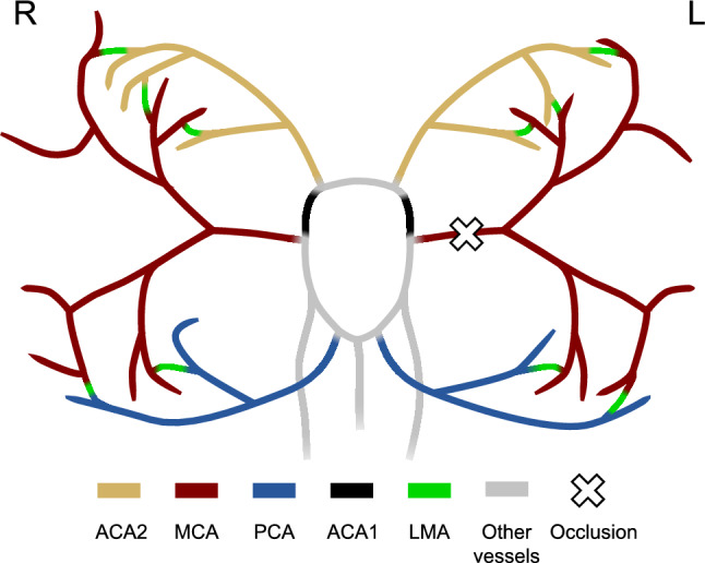
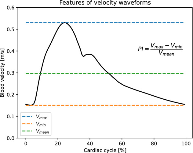
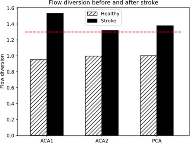
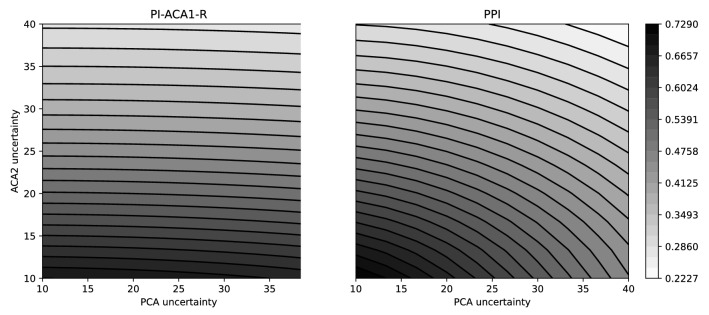
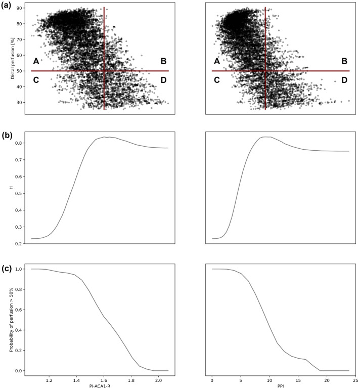
Assessment of distal cerebral perfusion after ischaemic stroke is currently only possible through expensive and time-consuming imaging procedures which require the injection of a contrast medium. Alternative approaches that could indicate earlier the impact of blood flow occlusion on distal cerebral perfusion are currently lacking. The aim of this study was to identify novel biomarkers suitable for clinical implementation using less invasive diagnostic techniques such as Transcranial Doppler (TCD). We used 1D modelling to simulate pre- and post-stroke velocity and flow wave propagation in a typical arterial network, and Sobol's sensitivity analysis, supported by the use of Gaussian process emulators, to identify biomarkers linked to cerebral perfusion. We showed that values of pulsatility index of the right anterior cerebral artery > 1.6 are associated with poor perfusion and may require immediate intervention. Three additional biomarkers with similar behaviour, all related to pulsatility indices, were identified. These results suggest that flow pulsatility measured at specific locations could be used to effectively estimate distal cerebral perfusion rates, and ultimately improve clinical diagnosis and management of ischaemic stroke.

Keywords: Biomarker; Brain circulation; Cardiovascular modelling; Gaussian process emulator; Ischaemic stroke; Leptomeningeal collateral; Sensitivity analysis; Wave propagation.


Figures

Figure 1
Figure 1
Portion of the network used in this study. ACA2 district is shown in yellow, MCA district in red, PCA district in blue, ACA1 in black, LMAs in green, other intracranial vessels in grey. Extracranial vessels are not shown. The cross identifies the occlusion location in the left MCA. The white circle indicates the locations where the velocities are measured to compute the biomarkers.
Figure 2
Figure 2
Typical velocity waveform from healthy ACA1 (black). Blue dotted line indicates its maximum value (peak systole), while orange and green dotted lines indicate the minimum(end-diastole) and time-averaged values respectively. The definition of pulsatility index (PI) for this waveform is shown on the right. The flow diversion (FD), not shown here, is computed as the ratio of the ipsilateral and contralateral average velocities.
Figure 3
Figure 3
The flow diversion in ACA1, ACA2 and PCA increases after stroke (black bars) with respect to the healthy case (hatched bars) and reaches values above 1.3 (red dashed line), which is a sign of LMA collateralisation.
Figure 4
Figure 4
Following left MCA stroke (black bars) the pulsatility index of left ACA1, ACA2 and PCA decreases, while the right side is only minimally affected. White bars show the values in the healthy case, and the red dashed line indicates PI=1.2: values before 1.2 are signs of distal LMA collateralisation.
Figure 5
Figure 5
Sobol indices in case of 40% uncertainty on input parameters (on the x axis). “R0: Vx” indicates the radius of vessel Vx, “WK: R” is the resistive part of the windkessel element. Darker colours signify higher influence of the inputs on the outputs (on the y axis). The average perfusion is significantly influenced by the radius of LMAs. With high level of uncertainty on the inputs the biomarkers do not depend on R0: LMA and thus cannot be used as proxy measurements.
Figure 6
Figure 6
Sobol indices in case of 10% uncertainty on left and right R0: ACA2 and R0: PCA. Uncertainty on remaining parameters is 40%. Increased knowledge of ACA2 and PCA radii causes an increase in the dependency of the biomarkers on the LMA radii. The four biomarkers whose Sobol index with R0: LMA is above 0.5 are highlighted.
Figure 7
Figure 7
Contour plots of correlation surfaces for biomarkers PI-ACA1-R (left) and PPI (right), obtained with uncertainties on radii of PCA and ACA2 ranging from 10% to 40%.
Figure 8
Figure 8
(a) Scatter plot of the distal perfusion as a function of the biomarkers. The perfusion is represented as a percentage of the healthy case. The biomarker PI-ACA1-R is on the left, while PPI on the right. The red horizontal line indicates a perfusion level of 50%, red vertical line indicates the biomarker threshold. Patients in A do not need immediate intervention and are classified correctly. Patients in B do not need immediate intervention and are classified incorrectly. Patients in C need immediate intervention and are classified incorrectly. Patients in D need immediate intervention and are classified correctly. (b) H ratio for biomarkers PI-ACA1-R (left) and PPI (right). The value of the biomarker that maximises H is chosen as the biomarker threshold. (c) Probability of perfusion >50% as a function of the observed biomarkers. Left: PI-ACA1-R. Right: PPI.

References

    1. Alastruey J, Parker KH, Peiro J, Byrd SM, Sherwin SJ. Modelling the circle of Willis to assess the effects of anatomical variations and occlusions on cerebral flows. J. Biomech. 2007;40(8):1794–1805. doi: 10.1016/j.jbiomech.2006.07.008. - DOI - PubMed
    1. Bang OY, Saver JL, Kim SJ, Kim GM, Chung CS, Ovbiagele B, et al. Collateral flow predicts response to endovascular therapy for acute ischemic stroke. Stroke. 2011;42(3):693–699. doi: 10.1161/STROKEAHA.110.595256. - DOI - PMC - PubMed
    1. Bellner, J., B. Romner, P. Reinstrup, K. A. Kristiansson, E. Ryding, L. Brandt. Transcranial Doppler sonography pulsatility index (PI) reflects intracranial pressure (ICP). Surg Neurol 62(1):45–51 (2004) - PubMed
    1. Benemerito, I., A. P. Narata, A. Narracott, A. Marzo. 1D model of the human cerebral vasculature: Circle of Willis and leptomeningeal anastomoses. Figshare. 202210.15131/shef.data.19119509
    1. Bertaglia G, Navas-Montilla A, Valiani A, Monge Garcia MI, Murillo J, Caleffi V. Computational hemodynamics in arteries with the one-dimensional augmented fluid-structure interaction system: viscoelastic parameters estimation and comparison with in-vivo data. J. Biomech. 2020;100:109595. doi: 10.1016/j.jbiomech.2019.109595. - DOI - PubMed