. 2022 Jun;30(3):919-934.
doi: 10.1007/s10787-022-00956-6. Epub 2022 Apr 1.

Neuroprotective effect of liraglutide in an experimental mouse model of multiple sclerosis: role of AMPK/SIRT1 signaling and NLRP3 inflammasome

Affiliations

Neuroprotective effect of liraglutide in an experimental mouse model of multiple sclerosis: role of AMPK/SIRT1 signaling and NLRP3 inflammasome

Reham A Ammar et al. Inflammopharmacology. 2022 Jun.

Abstract

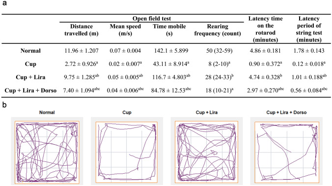
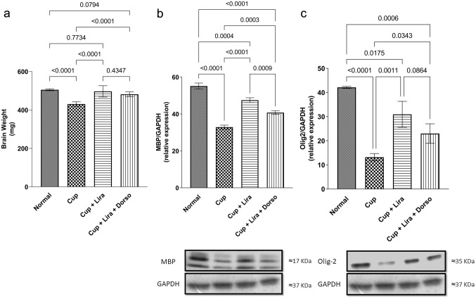
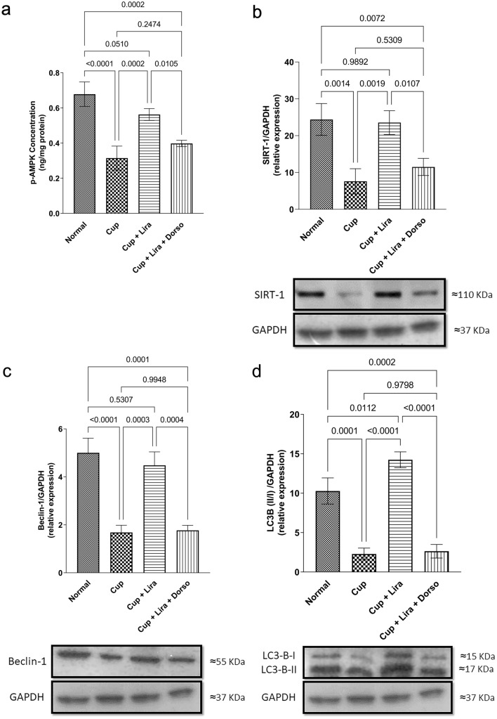
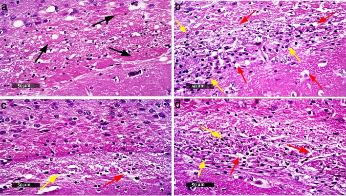
The heterogeneous nature of multiple sclerosis (MS) and the unavailability of treatments addressing its intricate network and reversing the disease state is yet an area that needs to be elucidated. Liraglutide, a glucagon-like peptide-1 analogue, recently exhibited intriguing potential neuroprotective effects. The currents study investigated its potential effect against mouse model of MS and the possible underlying mechanisms. Demyelination was induced in C57Bl/6 mice by cuprizone (400 mg/kg/day p.o.) for 5 weeks. Animals received either liraglutide (25 nmol/kg/day i.p.) or dorsomorphin, an AMPK inhibitor, (2.5 mg/Kg i.p.) 30 min before the liraglutide dose, for 4 weeks (starting from the second week). Liraglutide improved the behavioral profile in cuprizone-treated mice. Furthermore, it induced the re-myelination process through stimulating oligodendrocyte progenitor cells differentiation via Olig2 transcription activation, reflected by increased myelin basic protein and myelinated nerve fiber percentage. Liraglutide elevated the protein content of p-AMPK and SIRT1, in addition to the autophagy proteins Beclin-1 and LC3B. Liraglutide halted cellular damage as manifested by reduced HMGB1 protein and consequently TLR-4 downregulation, coupled with a decrease in NF-κB. Liraglutide also suppressed NLRP3 transcription. Dorsomorphin pre-administration indicated a possible interplay between AMPK/SIRT1 and NLRP3 inflammasome activation as it partially reversed liraglutide's effects. Immunohistochemical examination of Iba+ microglia emphasized these findings. In conclusion, liraglutide exerts neuroprotection against cuprizone-induced demyelination via anti-inflammatory, autophagic flux activation, NLRP3 inflammasome suppression, and anti-apoptotic mechanisms, possibly mediated, at least in part, via AMPK/SIRT1, autophagy, TLR-4/ NF-κB/NLRP3 signaling. The potential mechanistic insight of Lira in alleviating Cup-induced neurotoxicity via: (1) AMPK/SIRT1 pathways activation resulting in the stimulation of brain autophagy flux (confirmed by lowering Beclin-1 and LC3-B protein expression). (2) Inhibition of NLRP3 inflammasome activation, as evidenced by reduced HMGB1, TLR-4, NF-κB and NLRP3 protein expression, alongside diminishing the activation of its downstream cascade as reflected by reduced levels of caspase-1 and IL-1β protein expression. (3) A possible modulating interplay between the previously mentioned two pathways.

Keywords: AMPK/SIRT1; Autophagy; Dorsomorphin; Liraglutide; Multiple sclerosis; NLRP3 inflammasome.


Conflict of interest statement

The authors declare that there is no conflict of interest.

Figures

Fig. 1
Fig. 1
Experimental time scale of Lira and Dorso in Cup-induced demyelination mouse model
Fig. 2
Fig. 2
Effect of Lira on Cup-induced alterations in locomotion & motor ability in C57Bl/6 mice through OFT, rotarod and string test. a Table of the behavioural analysis results. Each value represents the mean ± S.D. except for the rearing frequency, values represent the median and range of 12 mice in the open field arena. b Represents track plots showing the position of the centre of the mice in OFT test. Statistical significance between groups was detected using one-way ANOVA followed by Tukey–Kramer post hoc tests except for rearing frequency, which was analyzed using Kruskal–Wallis ANOVA followed by Dunn’s post hoc statistical tests. aSignificantly different from Normal group, bSignificantly different from Cup group, cSignificantly different from Cup + Lira group at P < 0.05
Fig. 3
Fig. 3
Effect of Lira on Cup-induced alterations in brain weight and brain myelination biomarkers of C57Bl/6 mice. Vertical bars represent the mean ± S.D. of 12 mice for a brain weight, as well as six mice for b MBP and c Olig2 relative protein expression. Values are statistically significant at P < 0.05 using one-way ANOVA followed by Tukey–Kramer post hoc tests
Fig. 4
Fig. 4
Representative images showing the effect of Lira on Cup-induced deterioration in myelinated nerve fibers percentage. LFB-stained brain corpus callosum coronal sections (magnification, × 50) of: a Normal control group, b Cup-, c Lira-, d Lira + Dorso-treated groups LFB-staining intensities and e Bar chart for the percentage of myelinated nerve fibres in six random corpus callosum fields/tissue section. Vertical bars represent the mean ± S.D. of each group. Values are statistically significant at P < 0.05 using one-way ANOVA followed by Tukey–Kramer post hoc tests
Fig. 5
Fig. 5
Effect of Lira on brain p-AMPK/SIRT1 and autophagy biomarkers in Cup-induced demyelination in C57Bl/6 mice. Vertical bars represent the mean ± S.D. of six mice for a p-AMPK level, b SIRT1, c Beclin-1 and d LC3B (II/I) relative protein expression. Values are statistically significant at P < 0.05 using one-way ANOVA followed by Tukey–Kramer post hoc tests
Fig. 6
Fig. 6
Effect of Lira on Cup-induced inflammation and apoptosis via NLRP3 inflammasome activation in C57Bl/6 mice. Vertical bars represent the mean ± S.D. of six mice for a HMGB1, b & c TLR-4, d NF-κB, e NLRP3, f caspase-1 and g IL-1β protein expression. Values are statistically significant at P < 0.05 using one-way ANOVA followed by Tukey–Kramer post hoc tests
Fig. 7
Fig. 7
Effect of Lira on histopathological alterations in Cup-induced neurotoxicity. Representative images of H&E stained brain corpus callosum sections of C57Bl/6 mice, (magnification, × 50). a Normal control group, b Cup-, c Lira- and d Lira + Dorso-treated groups. Black arrow indicates abnormal glial cells infiltrates, yellow arrow indicates astroglial and microglial cells infiltrates and red arrow indicates demyelination, vacuolization and axonal damage
Fig. 8
Fig. 8
Effect of Lira on Cup-induced microgliosis. Representative images of anti-Iba-1 Immunohistochemical staining of brain corpus callosum sections of C57Bl/6 mice (magnification, × 50). a Normal control group, b Cup-, c Lira-, d Lira + Dorso-treated groups, and e Box plot chart for the count of active microglial cells that positively express Iba-1 in six random corpus callosum fields per tissue section. Vertical bars represent the median and range of each group. ( +) represents the mean. Values are statistically significant at P < 0.05 using Kruskal–Wallis ANOVA followed by Dunn’s post hoc statistical tests

References

    1. Abbas T, Faivre E, Hölscher C. Impairment of synaptic plasticity and memory formation in GLP-1 receptor KO mice: interaction between type 2 diabetes and Alzheimer’s disease. Behav Brain Res. 2009;205:265–271. doi: 10.1016/j.bbr.2009.06.035. - DOI - PubMed
    1. Abd El Aziz AE, Sayed RH, Sallam NA, El Sayed NS. Neuroprotective effects of telmisartan and nifedipine against cuprizone-induced demyelination and behavioral dysfunction in mice: roles of NF-κB and Nrf2. Inflammation. 2021;44:1629–1642. doi: 10.1007/s10753-021-01447-6. - DOI - PubMed
    1. Agostini L, Martinon F, Burns K, et al. NALP3 forms an IL-1β-processing inflammasome with increased activity in Muckle-Wells autoinflammatory disorder. Immunity. 2004;20:319–325. doi: 10.1016/S1074-7613(04)00046-9. - DOI - PubMed
    1. Badawi GA, Abd El Fattah MA, Zaki HF, El Sayed MI. Sitagliptin and liraglutide reversed nigrostriatal degeneration of rodent brain in rotenone-induced Parkinson’s disease. Inflammopharmacology. 2017;25:369–382. doi: 10.1007/s10787-017-0331-6. - DOI - PubMed
    1. Birnbaum Y, Bajaj M, Yang HC, Ye Y. Combined SGLT2 and DPP4 inhibition reduces the activation of the Nlrp3/ASC inflammasome and attenuates the development of diabetic nephropathy in mice with type 2 diabetes. Cardiovasc Drugs Ther. 2018;32:135–145. doi: 10.1007/s10557-018-6778-x. - DOI - PubMed

LinkOut - more resources