. 2022 Apr 1;22(1):140.
doi: 10.1186/s12872-022-02582-0.

Study protocol: MyoFit46-the cardiac sub-study of the MRC National Survey of Health and Development

Affiliations

Study protocol: MyoFit46-the cardiac sub-study of the MRC National Survey of Health and Development

Matthew Webber et al. BMC Cardiovasc Disord. .

Abstract

Background: The life course accumulation of overt and subclinical myocardial dysfunction contributes to older age mortality, frailty, disability and loss of independence. The Medical Research Council National Survey of Health and Development (NSHD) is the world's longest running continued surveillance birth cohort providing a unique opportunity to understand life course determinants of myocardial dysfunction as part of MyoFit46-the cardiac sub-study of the NSHD.

Methods: We aim to recruit 550 NSHD participants of approximately 75 years+ to undertake high-density surface electrocardiographic imaging (ECGI) and stress perfusion cardiovascular magnetic resonance (CMR). Through comprehensive myocardial tissue characterization and 4-dimensional flow we hope to better understand the burden of clinical and subclinical cardiovascular disease. Supercomputers will be used to combine the multi-scale ECGI and CMR datasets per participant. Rarely available, prospectively collected whole-of-life data on exposures, traditional risk factors and multimorbidity will be studied to identify risk trajectories, critical change periods, mediators and cumulative impacts on the myocardium.

Discussion: By combining well curated, prospectively acquired longitudinal data of the NSHD with novel CMR-ECGI data and sharing these results and associated pipelines with the CMR community, MyoFit46 seeks to transform our understanding of how early, mid and later-life risk factor trajectories interact to determine the state of cardiovascular health in older age.

Trial registration: Prospectively registered on ClinicalTrials.gov with trial ID: 19/LO/1774 Multimorbidity Life-Course Approach to Myocardial Health- A Cardiac Sub-Study of the MCRC National Survey of Health and Development (NSHD).

Keywords: 4-dimensional flow; Cardiovascular health; Cardiovascular magnetic resonance; Electrocardiographic imaging; Life course risk factors; Myocardial tissue characterization; Perfusion; Risk trajectories; Subclinical myocardial dysfunction.


Conflict of interest statement

The authors declare that they have no competing of interests.

Figures

Fig. 1
Fig. 1
MyoFit46 study flowchart. BP, Blood pressure; CMR, cardiac magnetic resonance imaging; ECG, electrocardiogram; GP, general practitioner; MRI, magnetic resonance imaging; REDCap, research electronic data capture; PACS, picture archiving and communication system
Fig. 2
Fig. 2
High-throughput reusable ECGI vest for CMR. A The garment is embedded with 256 uniformly distributed dry electrodes that connect to g.HIamp for the recordings. B To ensure good skin contact of the dry electrodes an inflatable gilet is worn over the electrode vest for the duration of the recordings. This design allows for rapid montage onto and off the chest. After the recording is complete the inflatable jacket and electrode vest are removed leaving only the marker vest that the participant then wears into the CMR scanner. ECGI, electrocardiographic imaging. Other abbreviations as in Fig. 1
Fig. 3
Fig. 3
MyoFit46 CMR protocol. 2/3/4CH, 2/3/4 chamber; 4D, 4-dimensional; Ao, aorta; AVLAX, aortic valve long axis; AVSAX, aortic valve short axis; ECV, extracellular volume; HASTE, half-fourier single-shot turbo spin-echo; LGE, late gadolinium enhancement; LVLAX, left ventricular long axis; LVSAX, left ventricular short axis
Fig. 4
Fig. 4
Measuring aortic pulse wave velocity (PWV). A Free breathing gradient echo phase contrast cine of the ascending aorta at the level of the pulmonary artery bifurcation. B Aortic ‘candy cane’ used to pilot the aortic flow. Points 4 and 12 are highlighted to show the linear plane used for transection and for measuring 3D distance using our dedicated software. C Flow wave curves of both the ascending and descending aorta after normalization for peak flow which are used to measure the transit time. D PWV (m/s) is then calculated as 3D distance divided by transit time. 3D, 3-dimensional; PWV, pulse wave velocity
Fig. 5
Fig. 5
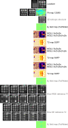
T1 and T2 Phantom QA framework. The same imaging protocol (defined here) is used for QA of multi-parametric T1 and T2 mapping data during the project lifecycle. Sequences in black are performed at baseline and then repeated every 8 weeks. Sequences in grey are performed at baseline and then for a total of 2 per year. 3D, 3-dimensional; FA, flip angle; IRSE, inversion recovery spin echo; MOLLI, modified Look-Locker inversion recovery; QA, quality assurance; SSFP, steady-state free precession
Fig. 6
Fig. 6
ECGI analysis workflow. A ECGI data is collected by g.Recorder software. B CMR image showing one transverse slice of the HASTE used to segment the epicardium. Note the fiducial MRI markers (white) situated on the anterior and posterior torso. C Ventricular segmentation of the same participant’s HASTE data, showing heart torso geometry and markers (Beekley medical, Bristol, Connecticut) following volume rendering, landmark localization and segmentation in Amira software (ThermoFisher, MA, USA, version 2020.3). D Signal averaging is performed using in house, customized software (Matlab, MathWorks, Natick, MA) and unipolar electrograms are then combined with Amira software heart torso geometry by solving the inverse problem of ECGI in collaboration with the Rudi laboratory. E Activation time isochrone maps in disease highlighting an area of fractionation corresponding to an area of LGE/scar in the mid anteroseptum of the left ventricle. EGM, electrogram. Other abbreviations as in Figs. 1, 2 and 3

References

    1. Roth GA, Mensah GA, Johnson CO, et al. Global burden of cardiovascular diseases and risk factors, 1990–2019: update from the GBD 2019 study. J Am Coll Cardiol. 2020;76(25):2982–3021. - PMC - PubMed
    1. Timmis A, Townsend N, Gale CP, et al. European society of cardiology: cardiovascular disease statistics 2019. Eur Heart J. 2020;41(1):12–85. - PubMed
    1. Abbafati C, Machado DB, Cislaghi B, et al. Global burden of 369 diseases and injuries in 204 countries and territories, 1990–2019: a systematic analysis for the Global Burden of Disease Study 2019. Lancet. 2020;396(10258):1204–1222. - PMC - PubMed
    1. Dai DF, Chen T, Johnson SC, Szeto H, Rabinovitch PS. Cardiac aging: from molecular mechanisms to significance in human health and disease. Antioxidants Redox Signal. 2012;16(12):1492–1536. - PMC - PubMed
    1. Pugh KG, Wei JY. Clinical implications of physiological changes in the aging heart. Drugs Aging. 2001;18(4):263–276. - PubMed

Publication types