Review
. 2021 Dec 18;28(4):513-543.
doi: 10.3390/pathophysiology28040034.

The Role of Exposomes in the Pathophysiology of Autoimmune Diseases I: Toxic Chemicals and Food

Affiliations
Review

The Role of Exposomes in the Pathophysiology of Autoimmune Diseases I: Toxic Chemicals and Food

Aristo Vojdani et al. Pathophysiology. .

Abstract

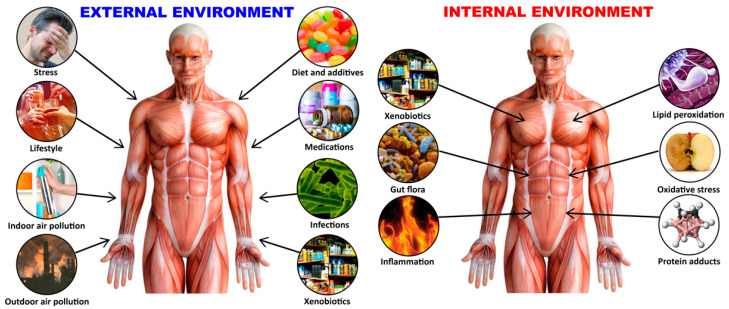
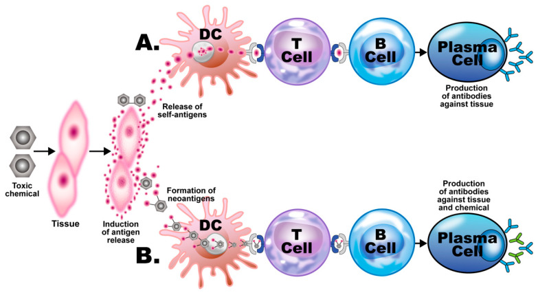
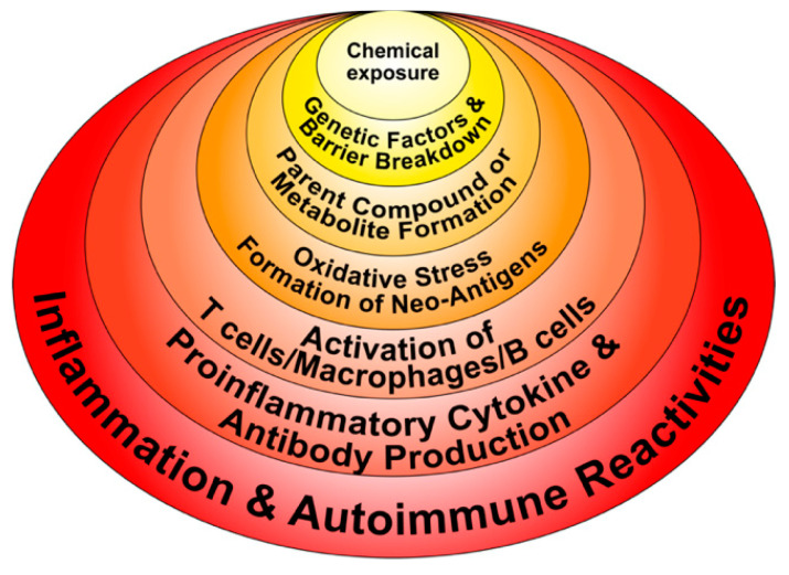
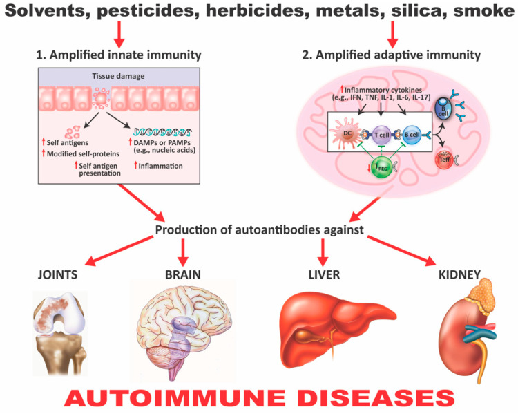
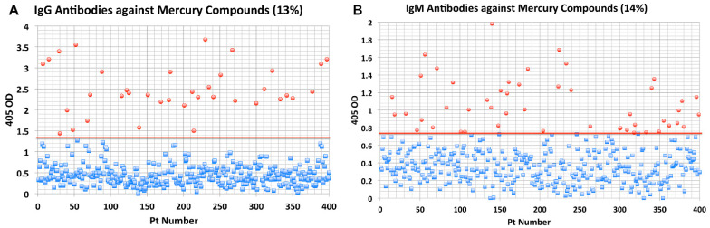
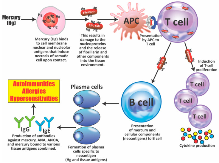
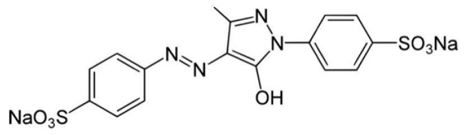
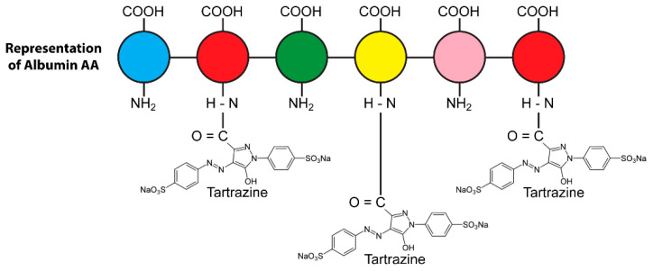
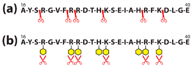
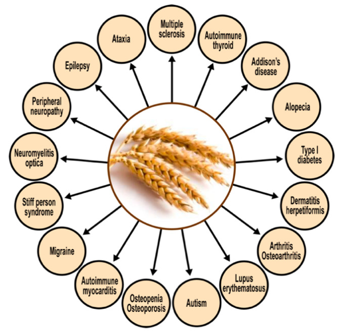
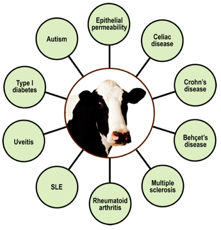
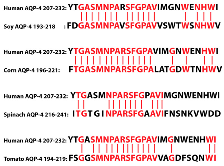
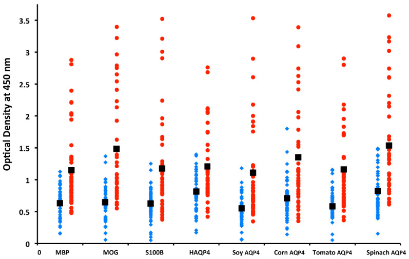
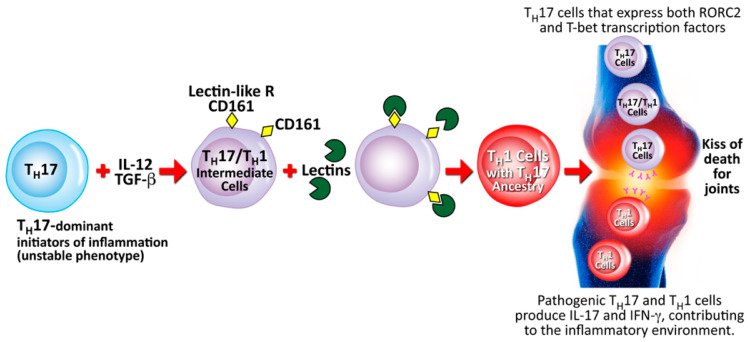
Autoimmune diseases affect 5-9% of the world's population. It is now known that genetics play a relatively small part in the pathophysiology of autoimmune disorders in general, and that environmental factors have a greater role. In this review, we examine the role of the exposome, an individual's lifetime exposure to external and internal factors, in the pathophysiology of autoimmune diseases. The most common of these environmental factors are toxic chemicals, food/diet, and infections. Toxic chemicals are in our food, drink, common products, the air, and even the land we walk on. Toxic chemicals can directly damage self-tissue and cause the release of autoantigens, or can bind to human tissue antigens and form neoantigens, which can provoke autoimmune response leading to autoimmunity. Other types of autoimmune responses can also be induced by toxic chemicals through various effects at the cellular and biochemical levels. The food we eat every day commonly has colorants, preservatives, or packaging-related chemical contamination. The food itself may be antigenic for susceptible individuals. The most common mechanism for food-related autoimmunity is molecular mimicry, in which the food's molecular structure bears a similarity with the structure of one or more self-tissues. The solution is to detect the trigger, remove it from the environment or diet, then repair the damage to the individual's body and health.

Keywords: autoimmune disease; environmental factors; exposome; food; molecular mimicry; toxic chemicals.


Conflict of interest statement

A.V. is co-owner, CEO, and technical director of Immunosciences Lab., Inc. E.V. is owner of Regenera Medical.

Figures

Figure 1
Figure 1
The exposome is an individual’s lifetime exposure to a variety of external and internal factors.
Figure 2
Figure 2
Gene plus exposome factors that contribute to autoimmune diseases.
Figure 3
Figure 3
Putative mechanisms of chemical-induced autoimmunity. Toxic chemicals damage host tissue, releasing tissue self-antigens. (A) The self-antigens are picked up by dendritic cells (DCs) and presented to T cells, which present them to B cells, inducing them to develop into plasma cells, which produce antibodies against the host tissue. (B) The toxic chemical metabolites bind to the self-tissue antigens, forming neoantigens, which go through the same process of presentation and development of the B cells into plasma cells, which, in this case, produce antibodies against both the body tissue and the chemical.
Figure 4
Figure 4
Potential molecular mechanisms implicated in chemical-induced autoimmune reactivities.
Figure 5
Figure 5
How xenobiotics can induce autoimmunity. (1) Amplified innate immunity. Tissue damage caused by xenobiotics can lead to the presence of cellular components and other damage-associated molecular pattern molecules (DAMPs) or pathogen-associated molecular pattern molecules (PAMPs). It can also lead to the release of self- and modified self-antigens, the presentation of these self-antigens to nontolerant lymphocytes, and the induction of inflammation. (2) Amplified adaptive immunity. The effects already described lead to the engagement of toll-like receptors (TLRs) and other innate sensors, the production of proinflammatory cytokines, a decrease in Treg populations, an increase in autoreactive T- and B-cell populations, and the production of autoantibodies against various self-tissues, which can contribute to autoimmune diseases. IFN = interferon; TNF = tumor necrosis factor; IL = interleukin; Treg = regulatory T cell; Teff = effector T cell.
Figure 6
Figure 6
Results for mercury antibodies expressed as optical density (OD) at 405 nm in the form of scattergrams: (A) mercury immunoglobulin G (IgG); (B) mercury immunoglobulin M (IgM).
Figure 7
Figure 7
The pathophysiology of inflammatory and autoimmune responses induced by mercury exposure. Within minutes to hours at the site of injection, mercury (Hg) binds to cellular components and then induces necrosis of somatic cells; this results not only in cell death, but also in tissue damage, the release of lysosomal enzymes, and the proteolysis of self-proteins such as nuclear and nucleolar proteins, which then carry mercury, helping in the formation of neoantigens. These antigens or peptides are taken up by antigen-presenting cells (APCs) and then presented first to T cells, and then to B cells. Mercury exposure also induces the production of B-cell activating factors by APCs, leading to the proliferation of both T cells and B cells. Mercury also promotes the production of cytokines, such as IL-4, and a Th2 response, inducing B cells to become plasma cells, which produce immunoglobulin G (IgG) or immunoglobulin E (IgE) antibodies against mercury, nuclear and nucleolar antigens such as fibrillarin, and chromatin and other autoantigens. The binding of these antibodies to antigens after activation of the complement cascade and binding to C1Q may result in the deposition of immune complexes in the kidney and possibly the joints. SSA = Sjögren’s syndrome A; SSB = Sjögren’s syndrome B; ANA = antinuclear antibodies; ANOA = antinucleolar antibodies.
Figure 8
Figure 8
Molecular formula for Tartrazine: C18H14N2Na2O8S2 (molecular weight 534.3).
Figure 9
Figure 9
Covalent binding of tartrazine through carboxylic group to human serum albumin amino groups, forming tartrazine–protein adduct.
Figure 10
Figure 10
Amino acid sequence of albumin peptide before (a) and after (b) food colorant binds to different amino acids contained in the chain. (a) Trypsin (symbolized by red scissors) is shown cleaving the amino acid chain of a 16–40 sequence of albumin; (b) colorants (symbolized by yellow hexagons) bind to the major amino acids arginine (R), histidine (H), and lysine (K) present in the albumin sequence, making it difficult for the trypsin to cleave the sequence, and decreasing digestive effectivity.
Figure 11
Figure 11
Spectrum of autoimmune disorders associated with wheat proteomes.
Figure 12
Figure 12
Spectrum of autoimmune disorders associated with milk proteomes. SLE = systemic lupus erythematosus.
Figure 13
Figure 13
Antigenic similarity between cow’s milk protein β-casein and islet cell autoantigen.
Figure 14
Figure 14
Similarity between cow’s milk protein BTN and human MOG. Only one out of many cross-reactive peptides is shown. MOG = myelin oligodendrocyte glycoprotein; BTN = butyrophilin.
Figure 15
Figure 15
Similarity between different plant AQP4s and human AQP4. AQP = aquaporin.
Figure 16
Figure 16
IgM antibody values of MS patients (red circles) versus controls (blue diamonds) for myelin basic protein (MBP), myelin oligodendrocyte glycoprotein (MOG), S100B, human aquaporin 4 (AQP4), and AQP4 for soy, corn, tomato, and spinach were significantly higher for patients than controls. IgG and IgA results showed similar differences between patients and controls.
Figure 17
Figure 17
Proposed mechanism for the contribution of lectins to the conversion of Th17 to Th17/Th1 and towards rheumatoid arthritis. TH = T-helper; IL = interleukin; TGF-β = transforming growth factor beta; CD = cluster of differentiation; ROR = retinoic acid-related orphan receptor; T-bet = T-box expressed in T cells; IFN-γ = interferon gamma.

References

    1. Opdenakker G., Abu El-Asrar A., Van Damme J. Remnant epitopes generating autoimmunity: From model to useful paradigm. Trends Immunol. 2020;41:367–378. doi: 10.1016/j.it.2020.03.004. - DOI - PubMed
    1. Tai Y., Wang Q., Korner H., Zhang L., Wei W. Molecular mechanisms of T cells activation by dendritic cells in autoimmune diseases. Front. Pharmacol. 2018;9:642. doi: 10.3389/fphar.2018.00642. - DOI - PMC - PubMed
    1. Steinman R.M. Lasker basic medical research award. Dendritic cells versatile controllers of the immune system. Nat. Med. 2007;13:1155–1159. doi: 10.1038/nm1643. - DOI - PubMed
    1. Lebre M.C., Jongbloed S.L., Tas S.W., Smeets T.J., Mcinnes I.B., Tak P.P. Rheumatoid arthritis synovium contains two subsets of CD83-DC-LAMP-dendritic cells with distinct cytokine profiles. Am. J. Pathol. 2008;172:940–950. doi: 10.2353/ajpath.2008.070703. - DOI - PMC - PubMed
    1. Tournadre A., Leniet V., Eljaafari A., Miossec P. Immature muscle precursors are a source of interferon-beta in myositis: Role of Toll-like receptor 3 activation and contribution to HLA class I up-regulation. Arthritis Rheum. 2012;64:533–541. doi: 10.1002/art.33350. - DOI - PubMed

LinkOut - more resources