. 2021 Jan 8;28(1):34-49.
doi: 10.3390/pathophysiology28010004.

Is an Immunosuppressive Microenvironment a Characteristic of Both Intra- and Extraparenchymal Central Nervous Tumors?

Affiliations

Is an Immunosuppressive Microenvironment a Characteristic of Both Intra- and Extraparenchymal Central Nervous Tumors?

Amina Soltani et al. Pathophysiology. .

Abstract

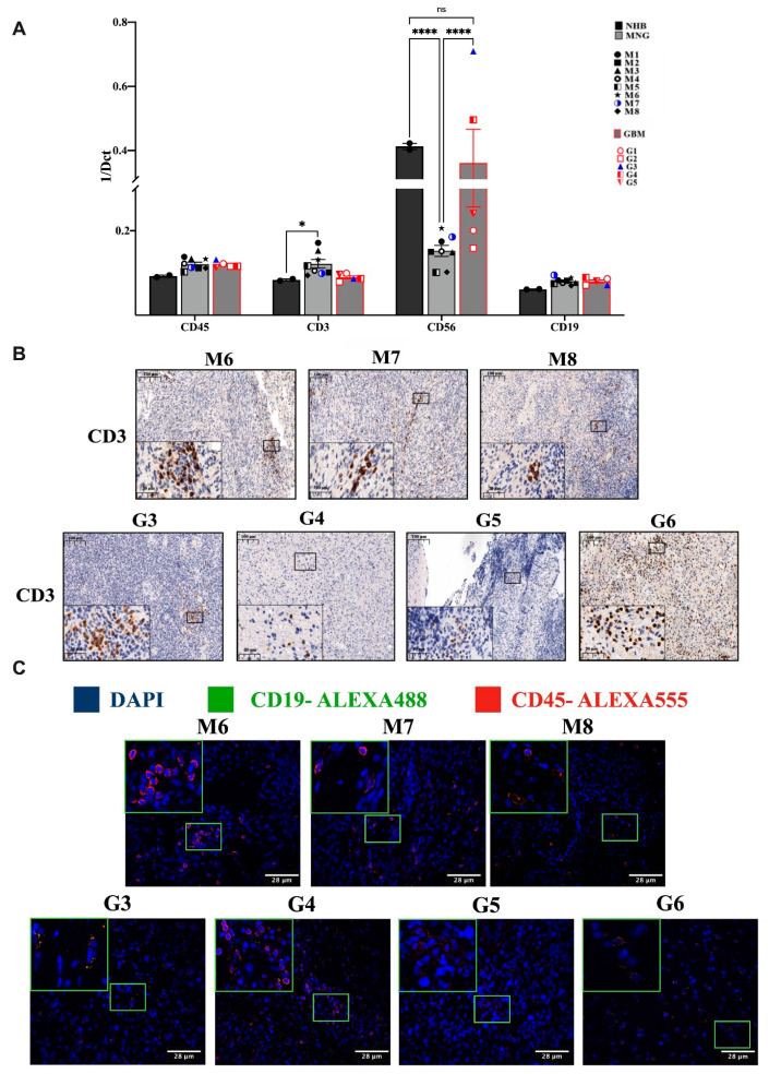
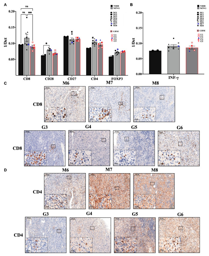
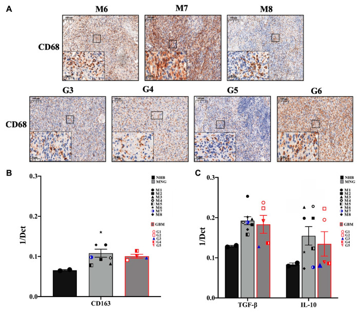
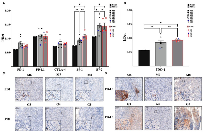
In spite of intensive research, the survival rates of patients diagnosed with tumors of the central nervous system (CNS) have not improved significantly in the last decade. Immunotherapy as novel and efficacious treatment option in several other malignancies has failed in neuro-oncology likely due to the immunosuppressive property of the brain tissues. Glioblastoma (GBM) is the most aggressive malignant CNS neoplasm, while meningioma (MNG) is a mainly low grade or benign brain tumor originating from the non-glial tissues of the CNS. The aim of the current preliminary study is to compare the immune microenvironment of MNG and GBM as potential target in immunotherapy. Interestingly, the immune microenvironment of MNG and GBM have proved to be similar. In both tumors types the immune suppressive elements including regulatory T cells (Treg), tumor-associated macrophages (TAM) were highly elevated. The cytokine environment supporting Treg differentiation and the presence of indoleamine 2,3-dioxygenase 1 (IDO1) have also increased the immunosuppressive microenvironment. The results of the present study show an immune suppressive microenvironment in both brain tumor types. In a follow-up study with a larger patient cohort can provide detailed background information on the immune status of individual patients and aid selection of the best immune checkpoint inhibitor or other immune modulatory therapy. Immune modulatory treatments in combination with IDO1 inhibitors might even become alternative therapy for relapsed, multiple and/or malignant MNG or chemo-resistant GBM.

Keywords: glioblastoma; immune microenvironment; immune suppression; meningioma.


Conflict of interest statement

The authors declare no conflict of interest.

Figures

Figure 1
Figure 1
Infiltration of immune cell populations into MNG and GBM. (A) mRNA levels of the general leukocyte antigen CD45 and general immune cell subpopulation CD3+ T, CD56+ NK and CD19+ B cell markers in MNG (n = 8) and GBM (n = 5). Data are presented as 1/dCt individually and as average ± SEM. Significant changes are marked as * and **** (p < 0.05 and p < 0.0001, respectively). (B) Immunohistochemistry staining of CD3 T-cell population in both brain tumor types (GBM and MNG), magnification ×20 and ×40, size bar 100 and 20 μm respectively. (C) Immunofluorescence staining of the general leucocytes marker CD45 and of CD19 in MNG (n = 3) and GBM (n = 4), magnification ×40, size bar 28 μm. Only red staining can be detected in MNG and yellow (overlapping red and green) in some GBM samples.
Figure 2
Figure 2
Evaluation of the immune profile of MNG and GBM. (A) mRNA expression levels of CD8 cytotoxic T-cells and CD4 T-helper cells, CD28 as costimulatory signal transducer for T-cells survival and activation, as well as FOXP3 marker characteristic for regulatory T cells. (B) The anti-tumor interferon-gamma (INF-γ) mRNA expression levels were evaluated in both brain tumor microenvironments. Data are presented 1/dCt individually and as average ± SEM. Significant changes are marked as * and ** (p < 0.05 and p < 0.001, respectively). (C) Immunohistochemistry staining of CD8 was performed in MNG (n = 3) and GBM (n = 4) samples, magnification ×20 and ×40, size bar 100 and 20 μm respectively. (D) Immunohistochemistry staining of CD4 cells in MNG and GBM, magnification ×20 and ×40, size bar 100 and 20 μm respectively.
Figure 3
Figure 3
Tumor-associated macrophages (TAM) and immunosuppressive cytokine expressions in MNG and in GBM. (A) Immunohistochemistry staining of CD68 indicate the higher protein expression of TAM in both tumors, magnification ×20 and ×40, size bar 100 and 20 μm respectively. (B) CD163 m-RNA expression levels which represents TAM marker in MNG and GBM. (C) mRNA expression levels of anti-inflammatory cytokines IL10 and transforming growth factor β (TGFβ) in MNG and GBM samples. Data are presented as 1/dCt individually and as average ± SEM. Significant changes are marked as * (p < 0.05).
Figure 4
Figure 4
Immune checkpoint molecules that mediate immune therapy response in MNG and GBM. (A) mRNA expression levels of PD-1, PD-L1, CTLA-4, B7-1, and B7-2 in MNG and GBM patient samples. (B) mRNA expression levels of Indoleamine 2,3-dioxygenase-1 (IDO-1) were tested in MNG and GBM. Data are presented as 1/dCt individually and as average ± SEM. Significant changes are marked as * (p < 0.05). (C,D) Both MNG and GBM samples were stained for PD-1 and PD-L1, magnification ×20 and ×40, size bar 100 and 20 μm respectively.

Similar articles

Cited by

References

    1. Siegel R.L., Miller K.D.M., Jemal A. Cancer statistics, 2018. CA Cancer J. Clin. 2018;68:7–30. doi: 10.3322/caac.21442. - DOI - PubMed
    1. Ilkhanizadeh S., Lau J., Huang M., Foster D.J., Wong R., Frantz A., Wang S., Weiss W.A., Persson A.I. Glial Progenitors as Targets for Transformation in Glioma. Adv. Cancer Res. 2014;121:1–65. doi: 10.1016/b978-0-12-800249-0.00001-9. - DOI - PMC - PubMed
    1. Gieryng A., Pszczolkowska D., Walentynowicz K.A., Rajan W.D., Kaminska B. Immune microenvironment of gliomas. Lab. Investig. 2017;97:498–518. doi: 10.1038/labinvest.2017.19. - DOI - PubMed
    1. Agarwala S.S., Kirkwood J.M. Temozolomide, a Novel Alkylating Agent with Activity in the Central Nervous System, May Improve the Treatment of Advanced Metastatic Melanoma. Oncologist. 2000;5:144–151. doi: 10.1634/theoncologist.5-2-144. - DOI - PubMed
    1. Harmancı A.S., Youngblood M.W., Clark V.E., Coşkun S., Henegariu O., Duran D., Erson-Omay E.Z., Kaulen L.D., Lee T.I., Abraham B.J., et al. Integrated genomic analyses of de novo pathways underlying atypical meningiomas. Nat. Commun. 2017;8:14433. doi: 10.1038/ncomms14433. - DOI - PMC - PubMed