Review
. 2021 Mar 10;28(1):155-188.
doi: 10.3390/pathophysiology28010011.

Pathophysiology of Lung Disease and Wound Repair in Cystic Fibrosis

Affiliations
Review

Pathophysiology of Lung Disease and Wound Repair in Cystic Fibrosis

Massimo Conese et al. Pathophysiology. .

Abstract

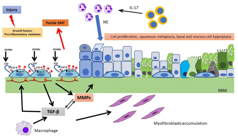
Cystic fibrosis (CF) is an autosomal recessive, life-threatening condition affecting many organs and tissues, the lung disease being the chief cause of morbidity and mortality. Mutations affecting the CF Transmembrane Conductance Regulator (CFTR) gene determine the expression of a dysfunctional protein that, in turn, triggers a pathophysiological cascade, leading to airway epithelium injury and remodeling. In vitro and in vivo studies point to a dysregulated regeneration and wound repair in CF airways, to be traced back to epithelial CFTR lack/dysfunction. Subsequent altered ion/fluid fluxes and/or signaling result in reduced cell migration and proliferation. Furthermore, the epithelial-mesenchymal transition appears to be partially triggered in CF, contributing to wound closure alteration. Finally, we pose our attention to diverse approaches to tackle this defect, discussing the therapeutic role of protease inhibitors, CFTR modulators and mesenchymal stem cells. Although the pathophysiology of wound repair in CF has been disclosed in some mechanisms, further studies are warranted to understand the cellular and molecular events in more details and to better address therapeutic interventions.

Keywords: CFTR; CFTR modulators; EGF/EGFR; airway epithelium; curcumin; cystic fibrosis; epithelial-mesenchymal transition; mesenchymal stem cells; wound healing.


Conflict of interest statement

The authors declare no conflict of interest.

Figures

Figure 1
Figure 1
Airway epithelial damage in the airways following infection with P. aeruginosa. (a) Hematoxylin and eosin-stained lung section from control mice and (b,c) hematoxylin and eosin-stained lung sections from P. aeruginosa infected mice 48 h post-intratracheal instillation at a dose of 1 × 10⁵ colony forming units. Panel c is an enlargement of panel b. Original magnification ×20 (a,b) and ×40 (c). White arrow in (c) indicates loss of the airway epithelium.
Figure 2
Figure 2
Wound repair and regeneration in a non-CF airway epithelium. Following injury, many cytokines and growth factors (e.g., EGF, HGF, FGF) are secreted in the wound repair microenvironment, which incite in basal cells a migratory and proliferative phenotype. Shedding of EGFR ligands (e.g., EGF) by ADAMs and binding to EGFR in an autocrine or iuxtacrine manner are key events involved in stimulation of cell migration and proliferation. Afterwards, MMP secretion by regenerating epithelial cells and subsequent TGF-β activation lead to genetic expression changes, related to EMT stimulation and activation of EMT-transcription factors (EMT-TFs). Epithelial cells and macrophages release TGF-β that induce ECM component deposition by epithelial cells and stimulate fibroblast activation, resulting in further matrix deposition. These events provoke alterations in junctional complexes and reorganization of actin cytoskeleton (not shown), modification of various integrin expression with β1-integrins increase at basal side and ectopic expression on the apical side, deposition of inflammatory ECM glycoproteins (e.g., fibronectin is shown) and its remodeling exerted by MMPs. TGF-β and MMPs enhance each other in a positive way. This process proceeds with the formation of a squamous stratified epithelium and subsequent pseudostratification and mucociliary differentiation.
Figure 3
Figure 3
The “air-opened” nude mouse xenograft model. Dissociated airway epithelial cells are seeded into the lumen of a denuded rat trachea tied at their end to sterile tubing. The assembly is subcutaneously implanted in the flank of a recipient nude mouse. Epithelial repair and regeneration steps result in the generation of a well-differentiated mucociliary epithelium (inset).
Figure 4
Figure 4
Methods of wound injury in different models of airway epithelial cell cultures. (a) Mechanical injury by the pipette tip (scratch assay). (b) Circular lesion produced by lethal electroporation. (c) Circular wounds obtained by an airbrush linked to a pressure regulator. (d) Submerged cultures of immortalized cell lines on plastic. (e) Polarized cultures of immortalized and primary cells seeded onto semipermeable filters. (f) ALI cultures on semipermeable filters. While immortalized cell lines polarize but not differentiate at ALI (e.g., Calu-3), primary airway epithelial cells differentiate at ALI into a pseudostratified epithelium presenting ciliated, basal, secretory, and mucus-producing goblet cells.
Figure 5
Figure 5
Wound repair and regeneration in a CF airway epithelium. The pro-inflammatory milieu of the CF airways and the partial EMT state of CF airway epithelial cells (as represented by the detachment of a single cell from the epithelial sheet)) stimulate processes involved in regeneration and repair that are exaggerated in regard to a non-CF epithelium (as one can observe by the arrow thickness in comparison to Figure 2). However, the wound repair does not occur properly, eventually leading to persistence of cell proliferation, squamous metaplasia, and basal and mucus cell hyperplasia. The thickening of reticular basement membrane (RBM) is likely due to elevated TGF-β levels and enhanced myofibroblast differentiation and accumulation. Neutrophils infiltrate the CF airways following IL-17 secretion by T cells and produce mediators (e.g., NE) that retard the wound repair.

Similar articles

Cited by

References

    1. Elborn J.S. Cystic fibrosis. Lancet. 2016;388:2519–2531. doi: 10.1016/S0140-6736(16)00576-6. - DOI - PubMed
    1. Amaral M.D. Novel personalized therapies for cystic fibrosis: Treating the basic defect in all patients. J. Intern. Med. 2015;277:155–166. doi: 10.1111/joim.12314. - DOI - PubMed
    1. Marson F.A.L., Bertuzzo C.S., Ribeiro J.D. Classification of CFTR mutation classes. Lancet Respir. Med. 2016;4:e37–e38. doi: 10.1016/S2213-2600(16)30188-6. - DOI - PubMed
    1. De Boeck K., Amaral M.D. Progress in therapies for cystic fibrosis. Lancet Respir. Med. 2016;4:662–674. doi: 10.1016/S2213-2600(16)00023-0. - DOI - PubMed
    1. Boucher R.C. Cystic fibrosis: A disease of vulnerability to airway surface dehydration. Trends Mol. Med. 2007;13:231–240. doi: 10.1016/j.molmed.2007.05.001. - DOI - PubMed