

Corneal Confocal Microscopy in the Diagnosis of Small Fiber Neuropathy: Faster, Easier, and More Efficient Than Skin Biopsy?
- PMID: 35366285
- PMCID: PMC8954271
- DOI: 10.3390/pathophysiology29010001
Corneal Confocal Microscopy in the Diagnosis of Small Fiber Neuropathy: Faster, Easier, and More Efficient Than Skin Biopsy?
Abstract
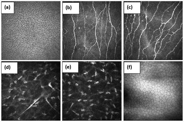
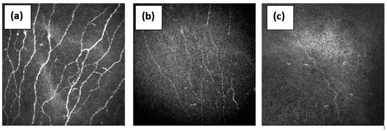
Chronic pain may affect 30-50% of the world's population and an important cause is small fiber neuropathy (SFN). Recent research suggests that autoimmune diseases may be one of the most common causes of small nerve fiber damage. There is low awareness of SFN among patients and clinicians and it is difficult to diagnose as routine electrophysiological methods only detect large fiber abnormalities, and specialized small fiber tests, like skin biopsy and quantitative sensory testing, are not routinely available. Corneal confocal microscopy (CCM) is a rapid, non-invasive, reproducible method for quantifying small nerve fiber degeneration and regeneration, and could be an important tool for diagnosing SFN. This review considers the advantages and disadvantages of CCM and highlights the evolution of this technique from a research tool to a diagnostic test for small fiber damage, which can be a valuable contribution to the study and management of autoimmune disease.
Keywords: Sjogren’s syndrome; autoimmune neuropathies; autoimmunity; confocal microscopy (CM); cornea; sarcoidosis; skin biopsy; small fiber neuropathy (SFN).
Conflict of interest statement
The authors declare no conflict of interest.
Figures
References
-
- Basantsova N.Y., Zinchenko Y.U.S., Starshinova A.A., Yablonsky P.K. Features of the diagnosis of small fiber neuropathies in various diseases (literature review) Pediatrician. 2018;9:101–110. doi: 10.17816/PED96101-110. - DOI
-
- Johnson S.A., Shouman K., Shelly S., Sandroni P., Berini S.E., Dyck P.J.B., Hoffman E.M., Mandrekar J., Niu Z., Lamb C.J., et al. Small Fiber Neuropathy Incidence, Prevalence, Longitudinal Impairments, and Disability. Neurology. 2021;97:e2236–e2247. doi: 10.1212/WNL.0000000000012894. - DOI - PMC - PubMed
Publication types
LinkOut - more resources
Full Text Sources
Research Materials
Miscellaneous