

Kimura's disease successively affecting multiple body parts: a case-based literature review
- PMID: 35366827
- PMCID: PMC8977031
- DOI: 10.1186/s12886-022-02378-y
Kimura's disease successively affecting multiple body parts: a case-based literature review
Abstract
Background: Kimura's disease is a rare, benign, chronic inflammatory disease that presents as painless, solid masses mainly affecting the deep subcutaneous areas of the head and neck, especially the salivary glands, parotid glands and nearby lymph nodes. It is characterized by elevated peripheral blood eosinophil and Immunoglobulin E (IgE) levels.
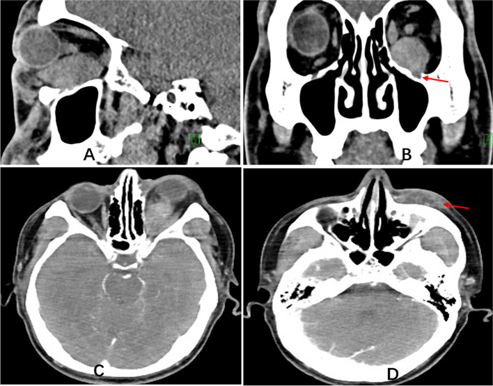
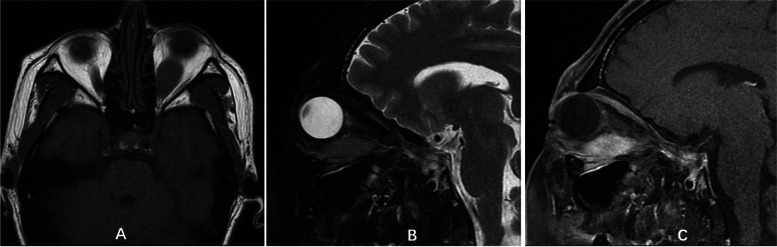
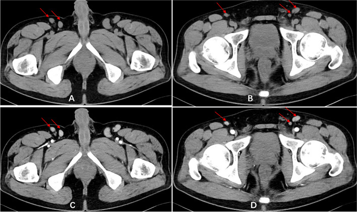
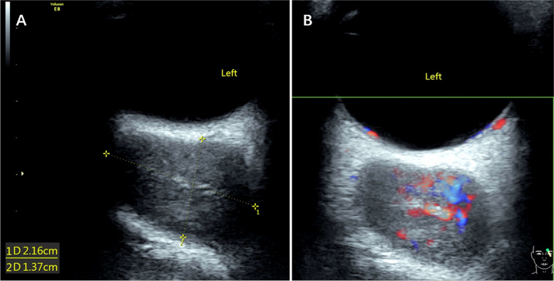
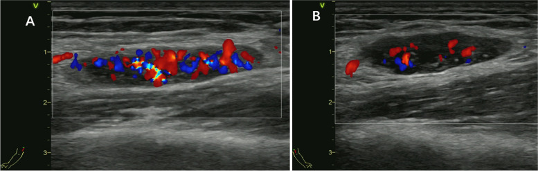
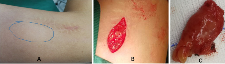
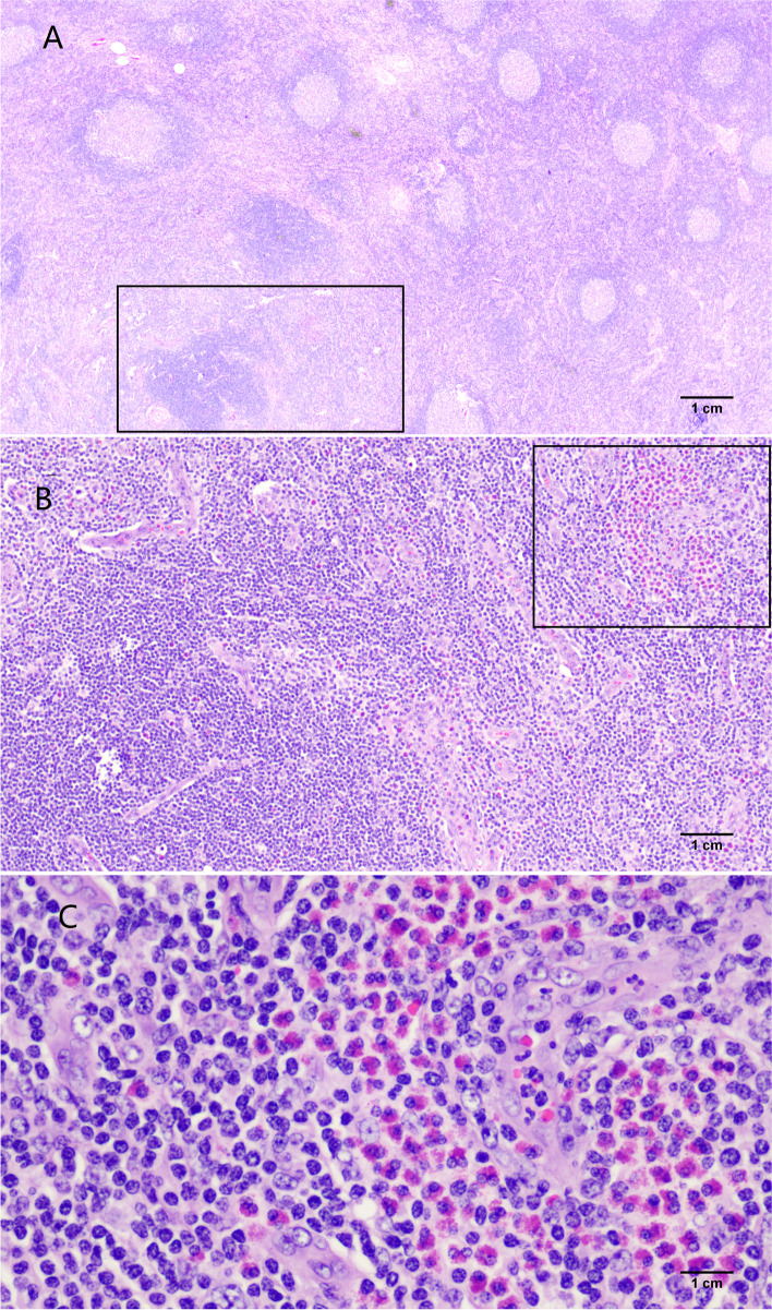
Case presentation: A 31-year-old Asian male presented with an orbital space-occupying lesion lasting for 1.5 years. Ten years prior, surgical excision of bilateral fossa cubitalis and groin masses was performed, and the pathological examination showed "lymphoproliferative disease". One year later, masses reappeared near the surgical sites; they grew slowly and shrank after glucocorticoid treatment. At this point, admission examinations showed in the peripheral blood an eosinophil proportion of 13.4%, a total IgE level of 26,900.00 IU/mL, prurigo present on the whole body, and multiple palpable masses near the bilateral fossa cubitalis and groin. The left eyeball was exophthalmic. The left elbow mass was excised, and the pathological examination confirmed Kimura's disease. Oral glucocorticoid therapy is taken and tapering regularly. The eosinophil count returned to normal, the IgE level gradually decreased, the orbital space-occupying lesion and elbow and groin masses shrank significantly, and the whole-body skin prurigo disappeared. Currently, the patient has been in a stable condition for eighteen months.
Conclusion: Our case provides a novel insight that Kimura's disease should be involved in the differential diagnosis of inflammatory lesion mass of orbit and also supports systemic regular glucocorticoid as a valuable therapy of such condition, but close follow-up and long-term observation are crucial.
Keywords: Eosinophils; Fossa Cubitalis; Groin; Immunoglobulin E; Kimura’s disease; Orbit.
© 2022. The Author(s).
Conflict of interest statement
All authors declare that they have no competing interests.
Figures

Similar articles
-
Kimura's Disease Diagnosed in the Department of Orthopedic Surgery Treated With Wide Excision: Report of Two Cases.In Vivo. 2023 May-Jun;37(3):1373-1378. doi: 10.21873/invivo.13219. In Vivo. 2023. PMID: 37103071 Free PMC article.
-
A common presentation of an uncommon pathology: Kimura disease.Trop Doct. 2023 Oct;53(4):512-516. doi: 10.1177/00494755231177487. Epub 2023 May 29. Trop Doct. 2023. PMID: 37248672
-
Kimura's disease sequentially involving multiple sites in the head and neck: A case report with a 13-year follow-up and literature review.J Int Med Res. 2025 May;53(5):3000605251337422. doi: 10.1177/03000605251337422. Epub 2025 May 13. J Int Med Res. 2025. PMID: 40357910 Free PMC article. Review.
-
Kimura's disease: a diagnostic challenge.Pediatrics. 2002 Sep;110(3):e39. doi: 10.1542/peds.110.3.e39. Pediatrics. 2002. PMID: 12205289
-
Kimura's disease presenting with intraparotid and neck nodes: A case report and review of literature.Ear Nose Throat J. 2025 Mar;104(1_suppl):326S-330S. doi: 10.1177/01455613221144495. Epub 2022 Dec 7. Ear Nose Throat J. 2025. PMID: 36476131 Review.
Cited by
-
Kimura disease in children: A report of 11 cases and review of the literature.Front Pediatr. 2023 Feb 17;11:1131963. doi: 10.3389/fped.2023.1131963. eCollection 2023. Front Pediatr. 2023. PMID: 36873634 Free PMC article.
-
Clinical results of definitive radiotherapy for local recurrent kimura disease in the head and neck after surgery: A retrospective study.Precis Radiat Oncol. 2024 Aug 21;8(3):119-125. doi: 10.1002/pro6.1238. eCollection 2024 Sep. Precis Radiat Oncol. 2024. PMID: 40336974 Free PMC article.
-
Kimura disease of the tongue base: a rare case diagnosed through cytological examination of Warthin-Finkeldey-type multinucleated cells.J Clin Exp Hematop. 2025 Jun 28;65(2):129-134. doi: 10.3960/jslrt.25007. Epub 2025 Mar 12. J Clin Exp Hematop. 2025. PMID: 40074363 Free PMC article.
-
Characteristics of 18F-FDG PET/CT in patients with Kimura's disease from China.BMC Med Imaging. 2024 Oct 8;24(1):269. doi: 10.1186/s12880-024-01446-7. BMC Med Imaging. 2024. PMID: 39379895 Free PMC article.
-
Dupilumab combined with corticosteroid therapy for Kimura disease with multiple systemic masses: a case report and literature review.Front Immunol. 2024 Oct 24;15:1492547. doi: 10.3389/fimmu.2024.1492547. eCollection 2024. Front Immunol. 2024. PMID: 39512341 Free PMC article. Review.
References
-
- Pitak-Arnnop P, Bellefqih S, Chaine A, Dhanuthai K, Bertrand JC, Bertolus C. Head and neck lesions of Kimura's disease: exclusion of human herpesvirus-8 and Epstein-Barr virus by in situ hybridisation and polymerase chain reaction. J Craniomaxillofac Surg. 2010;38(4):266–270. doi: 10.1016/j.jcms.2009.08.001. - DOI - PubMed
-
- Dik VK, van de Wiel BA, Vasmel WL. Kimura's disease of the parotid glands and multiple cervical lymph nodes. Neth J Med. 2010;68(4):175–177. - PubMed
Publication types
MeSH terms
LinkOut - more resources
Full Text Sources
