. 2022 Apr 2;18(1):126.
doi: 10.1186/s12917-022-03230-7.

NADH oxidase of Mycoplasma hyopneumoniae functions as a potential mediator of virulence

Affiliations

NADH oxidase of Mycoplasma hyopneumoniae functions as a potential mediator of virulence

Fei Hao et al. BMC Vet Res. .

Abstract

Background: Mycoplasma hyopneumoniae (M. hyopneumoniae) is the etiological agent of enzootic pneumonia, a highly infectious swine respiratory disease that distributed worldwide. The pathogenesis and virulence factors of M. hyopneumoniae are not fully clarified. As an important virulence factor of bacteria, nicotinamide adenine dinucleotide (NADH) oxidase (NOX) participates in host-pathogen interaction, however, the function of NOX involved in the pathogenesis of M. hyopneumoniae is not clear.

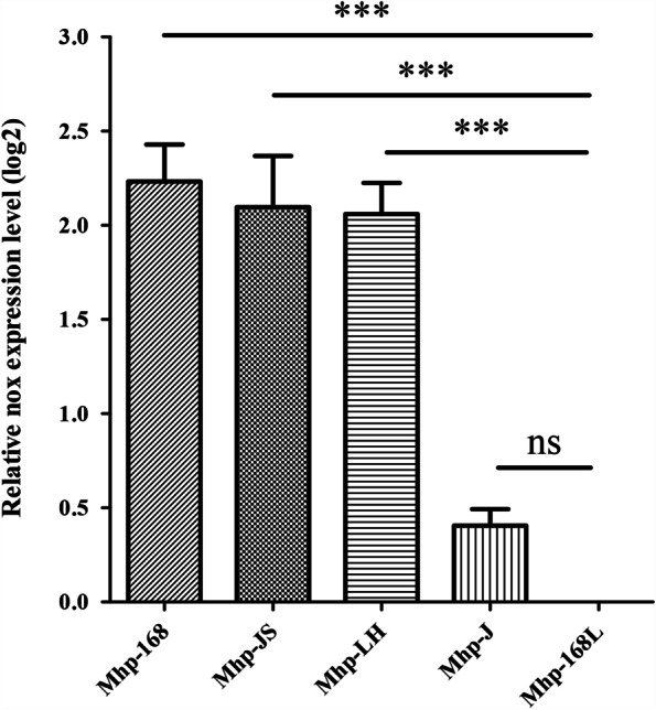
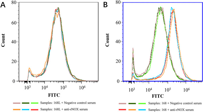
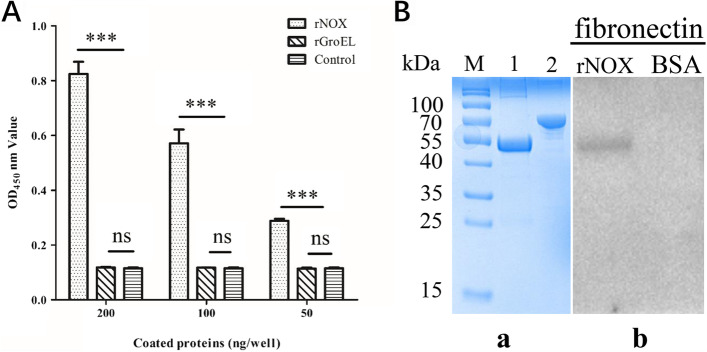
Results: In this study, significant differences in NOX transcription expression levels among different strains of M. hyopneumoniae differed in virulence were identified, suggesting that NOX may be correlated with M. hyopneumoniae virulence. The nox gene of M. hyopneumoniae was cloned and expressed in Escherichia coli, and polyclonal antibodies against recombinant NOX (rNOX) were prepared. We confirmed the enzymatic activity of rNOX based on its capacity to oxidize NADH to NAD+. Flow cytometry analysis demonstrated the surface localization of NOX, and subcellular localization analysis further demonstrated that NOX exists in both the cytoplasm and cell membrane. rNOX was depicted to mediate adhesion to immortalized porcine bronchial epithelial cells (hTERT-PBECs). Pre-neutralizing M. hyopneumoniae with anti-rNOX antibody resulted in a more than 55% reduction in the adhesion rate of high- and low-virulence M. hyopneumoniae strains to hTERT-PBECs. Moreover, a significant difference appeared in the decline in CCU50 titer between virulent (168) and virulence-attenuated (168L) strains. NOX not only recognized and interacted with host fibronectin but also induced cellular oxidative stress and apoptosis in hTERT-PBECs. The release of lactate dehydrogenase by NOX in hTERT-PBECs was positively correlated with the virulence of M. hyopneumoniae strains.

Conclusions: NOX is considered to be a potential virulence factor of M. hyopneumoniae and may play a significant role in mediating its pathogenesis.

Keywords: Adhesion; Mycoplasma hyopneumoniae; NADH oxidase; Virulence factor.


Conflict of interest statement

The authors declare that they have no competing interests.

Figures

Fig. 1
Fig. 1
Relative expression levels of nox genes involved in different M. hyopneumoniae (Mhp) compared to Mhp strain 168L (set to 1). Gene expression was determined by quantitative real-time PCR analysis. Error bars represent standard deviations from three independent experiments (***: p<0.001; ns: p >0.05)
Fig. 2
Fig. 2
Detection of surface-exposed NOX by flow cytometry. Comparison of fluorescence intensity of Mhp strains differed in virulence when treated with anti-NOX serum and negative serum. A: Negative control, Mhp strain 168L treated with pre-immune serum; Mhp strains 168L treated with anti-Mhp NOX serum. B: Negative control, Mhp strain 168 treated with pre-immune serum; Mhp strains 168 treated with anti-Mhp NOX
Fig. 3
Fig. 3
Subcellular localization of NADH oxidase in Mhp strains with different degrees of Virulence. Lane M is pre-stained protein mass markers, Lane 1, whole cell lysates of Mhp strain 168; Lane 2, cytoplasmic proteins of Mhp strain 168; Lane 3, membrane proteins of Mhp strain 168; Lane 4, whole cell lysates of Mhp strain 168L; Lane 5, cytoplasmic proteins of Mhp strain 168L; Lane 6, membrane proteins of Mhp strain 168L
Fig. 4
Fig. 4
rNOX adhesion assay with IFA. TRITC, antibody reactivity to IgG observed with TRITC-conjugated anti-rabbit IgG antibody (orange red); DAPI, nuclei of all cells were stained by DAPI reagent (blue); an overlay of the images is shown in the column labeled ‘‘Merge’’. Captured images were observed at 400× magnification. The red bar represents the scale: 100 μm
Fig. 5
Fig. 5
rNOX adhesive inhibition assays with real-time PCR and CCU50 assay. A Adhesion rates were calculated by real-time PCR for Mhp bacterial counting. Adhesion rate = (number of Mhp antigens collected from infected cells incubated with the anti-rNOX serum/ number of Mhp antigens collected from infected cells in the group incubated with the pre-immune sera) × 100. Quantitative real-time PCR analysis was performed and expressed as log10 DNA copy number per mL of Mhp strain-infected cell distribution against P97. Data are presented as the mean ± SD of three independent experiments (***: p<0.001). B Mhp titers were quantified using a CCU50 assay. Data are presented as the mean ± SD of three independent experiments (***: p<0.001; **: p<0.01).
Fig. 6
Fig. 6
A rNOX, rGroEL, and PBS were applied to examine fibronectin activity with indirect ELISA. *** represents an extremely significant difference (p < 0.001), while “ns” represents no significant difference between groups (p >0.05). B “a,” SDS–PAGE analysis of purified recombinant protein rNOX and BSA. Lane M is pre-stained protein mass markers; Lane 1: purified recombinant protein rNOX; Lane 2: BSA. The gel in this figure was cropped and the full-length gel was presented in Supplementary FigureS3. “b,” Mhp NOX with fibronectin interaction analysis by far Western blot. Lane rNOX: PVDF membrane with transferred Mhp NOX protein incubated with fibronectin and the anti-fibronectin antibody. Lane BSA: PVDF membrane with transferred BSA (negative control) incubated with fibronectin, and the anti-fibronectin antibody. The blots in this figure were cropped and the full-length blots were presented in Supplementary FigureS4.
Fig. 7
Fig. 7
Cytotoxicity and oxidative stress of hTERT-PBECs induced by rNOX. A CytoTox 96® Non-Radioactive Cytotoxicity Assay of hTERT-PBEC cytotoxicity after treatment with different concentrations of rNOX and various strains of Mhp with varying degrees of virulence (strains JS, 168, J and 168L). B ROS-Glo™ H2O2 assay of host cell cellular oxidative stress after treatment with rNOX and Mhp strains JS, 168, J and 168L
Fig. 8
Fig. 8
The apoptosis of hTERT-PBECs induced by rNOX and different virulent Mhp strains was assessed by flow cytometry using the annexin-V-FITC/PI double staining method 12 h post-infection. A: Negative control hTERT-PBECs cultured for 12 h in DMEM/F12 medium without FBS and growth factors before double staining; rNOX, the apoptosis rate of recombinant rNOX protein-infected hTERT-PBECs 12 h post-infection before double staining. B: Highly virulent Mhp strain 168 infected with hTERT-PBECs for 12 h treated without and with rNOX polyclonal antibody. C: Low virulent Mhp strain 168L infected with hTERT-PBECs for 12 h treated without and with rNOX polyclonal antibody

Similar articles

Cited by

References

    1. Opriessnig T, Gimenez-Lirola LG, Halbur PG. Polymicrobial respiratory disease in pigs. Anim Health Res Rev. 2011;12(2):133–48. - PubMed
    1. Maes D, Sibila M, Kuhnert P, Segales J, Haesebrouck F, Pieters M. Update on Mycoplasma hyopneumoniae infections in pigs: Knowledge gaps for improved disease control. Transbound Emerg Dis. 2018;65(Suppl 1):110–24. - PubMed
    1. Thacker EL, Thacker BJ, Janke BH. Interaction between Mycoplasma hyopneumoniae and swine influenza virus. J Clin Microbiol. 2001;39(7):2525–30. - PMC - PubMed
    1. Saade G, Deblanc C, Bougon J, Marois-Crehan C, Fablet C, Auray G, Belloc C, Leblanc-Maridor M, Gagnon CA, Zhu J, et al. Coinfections and their molecular consequences in the porcine respiratory tract. Vet Res. 2020;51(1):80. - PMC - PubMed
    1. Leal Zimmer FMA, Paes JA, Zaha A, Ferreira HB. Pathogenicity & virulence of Mycoplasma hyopneumoniae. Virulence. 2020;11(1):1600–22. - PMC - PubMed