

Does zinc oxide nanoparticles potentiate the regenerative effect of platelet-rich fibrin in healing of critical bone defect in rabbits?
- PMID: 35366880
- PMCID: PMC8976312
- DOI: 10.1186/s12917-022-03231-6
Does zinc oxide nanoparticles potentiate the regenerative effect of platelet-rich fibrin in healing of critical bone defect in rabbits?
Abstract
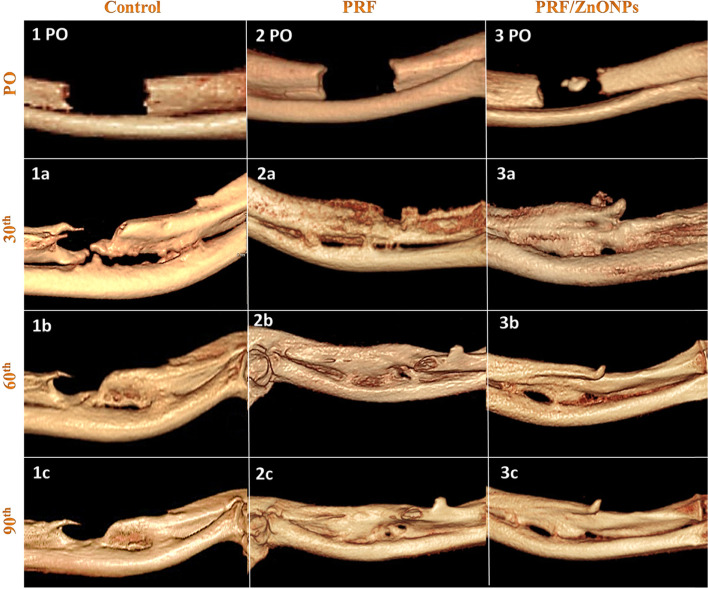
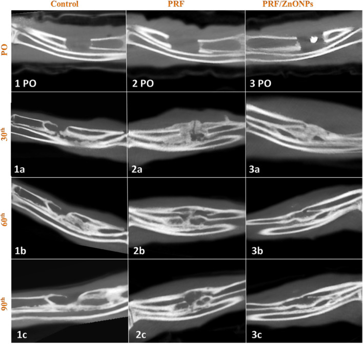
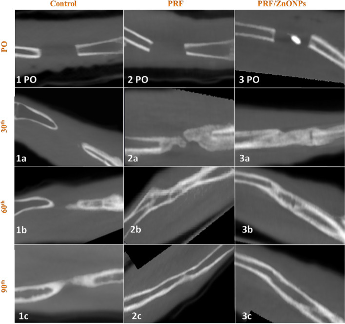
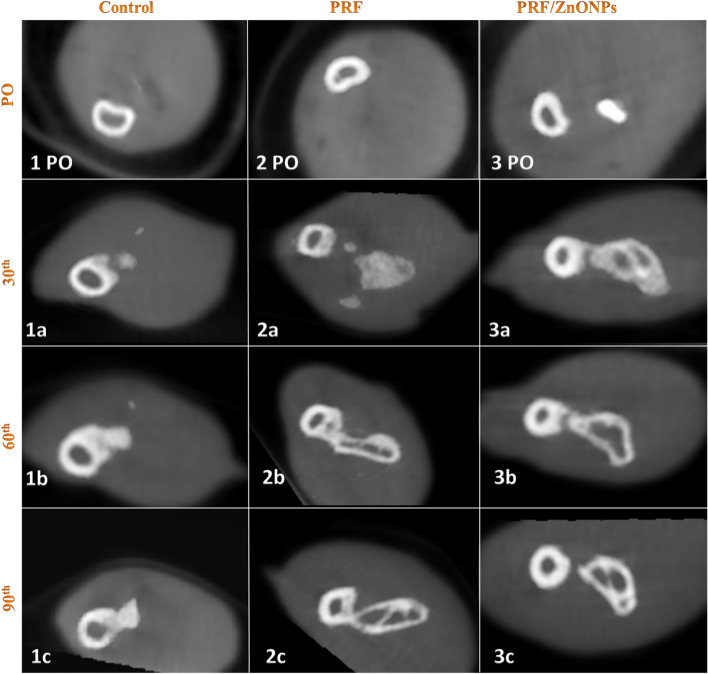
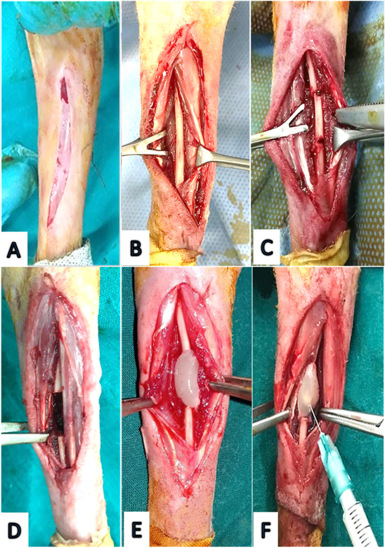
Background: Many encouraging studies confirmed the ability of Zinc Oxide Nanoparticles (ZnONPs) in accelerating bone growth and mineralization. The use of Platelet Rich-Fibrin (PRF) as a sole filling material for large segmental bone defects remains questionable. The objectives are to investigate the regenerative efficacy of autologous Platelet Rich-Fibrin (PRF) and Zinc Oxide Nanoparticles (ZnONPs) in repairing large segmental bone ulnar defects in a randomized controlled study in rabbits using computed tomographic interpretations. A 12 mm critical size defect was surgically induced in the ulna of 30 rabbits (n = 10/ group). In the control group, the defect was left empty. In the PRF group, the defect is filled with PRF. In the PRF/ZnONPs group, the defect is filled with PRF that was inoculated with 0.1 ml of 0.2% ZnONPs. Radiologic healing capacity was evaluated at the first, second, and third postoperative months.
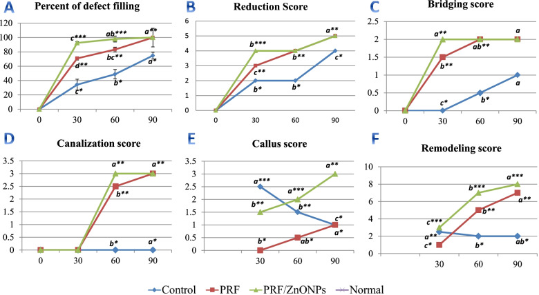
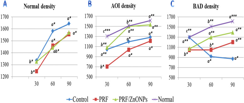
Results: Statistical analysis showed significant differences in the radiologic healing scores between the groups (P = 0.000-0.0001) at all-time points (P = 0.000-0.047) during the study.
Conclusion: Rabbits in the PRF/ZnONPs group showed the highest appreciable bone quality and quantity followed by the PRF group with high quantity but low bone quality meanwhile, rabbits in the control group showed minimal quantity but medium bone quality. Interestingly, the addition of ZnONPs to PRF can accelerate the healing of ulnar critical-size defects in rabbits.
Keywords: Bone density; Bridging; Canalization; Critical bone defect; Nanoparticles; Platelet-rich fibrin; Remodeling; Zinc oxide.
© 2022. The Author(s).
Conflict of interest statement
The authors declare that they have no competing interests.
Figures
Similar articles
-
Evaluation of the effects of platelet-rich fibrin on bone regeneration in diabetic rabbits.J Craniomaxillofac Surg. 2016 Feb;44(2):126-33. doi: 10.1016/j.jcms.2015.11.009. Epub 2015 Dec 8. J Craniomaxillofac Surg. 2016. PMID: 26732635
-
Comparative Analysis of Local Application of Titanium-Platelet Rich Fibrin (T-PRF) and Leukocyte-Platelet Rich Fibrin (L-PRF) in Bone Defect Healing: A Micro-CT and Histopathological Study in Rabbit Models.Microsc Res Tech. 2025 Jul;88(7):2073-2084. doi: 10.1002/jemt.24841. Epub 2025 Mar 6. Microsc Res Tech. 2025. PMID: 40047255 Free PMC article.
-
The impact of autogenous bone grafts on the regeneration of radial bone defects in rabbits compared to autogenous advanced platelet-rich fibrin plus.Open Vet J. 2025 Jan;15(1):325-338. doi: 10.5455/OVJ.2025.v15.i1.31. Epub 2025 Jan 31. Open Vet J. 2025. PMID: 40092204 Free PMC article.
-
Platelet-rich fibrin as a tissue engineering material in accelerate bone healing in rat bone defects: A systematic review and meta-analysis.Ann Med Surg (Lond). 2022 Nov 19;84:104869. doi: 10.1016/j.amsu.2022.104869. eCollection 2022 Dec. Ann Med Surg (Lond). 2022. PMID: 36504707 Free PMC article. Review.
-
Platelet-Rich Fibrin in Maxillary Sinus Augmentation: A Systematic Review.J Oral Implantol. 2015 Dec;41(6):746-53. doi: 10.1563/aaid-joi-D-14-00167. Epub 2014 Dec 23. J Oral Implantol. 2015. PMID: 25536095
Cited by
-
Significance and considerations of establishing standardized critical values for critical size defects in animal models of bone tissue regeneration.Heliyon. 2024 Jun 29;10(13):e33768. doi: 10.1016/j.heliyon.2024.e33768. eCollection 2024 Jul 15. Heliyon. 2024. PMID: 39071581 Free PMC article. Review.
-
Synergy of zinc oxide nanoparticles to losartan attenuates kidney injury induced by unilateral ureteral obstruction through modulation of the TNF-α/IL6 and BAX/BCL2 signaling pathways.Int Urol Nephrol. 2025 Jul;57(7):1989-2012. doi: 10.1007/s11255-024-04331-y. Epub 2025 Jan 14. Int Urol Nephrol. 2025. PMID: 39810058
-
Platelet-rich fibrin as an autologous biomaterial for bone regeneration: mechanisms, applications, optimization.Front Bioeng Biotechnol. 2024 Apr 16;12:1286035. doi: 10.3389/fbioe.2024.1286035. eCollection 2024. Front Bioeng Biotechnol. 2024. PMID: 38689760 Free PMC article. Review.
-
Navigating the combinations of platelet-rich fibrin with biomaterials used in maxillofacial surgery.Front Bioeng Biotechnol. 2024 Oct 7;12:1465019. doi: 10.3389/fbioe.2024.1465019. eCollection 2024. Front Bioeng Biotechnol. 2024. PMID: 39434715 Free PMC article. Review.
-
Bone Tissue Engineering and Nanotechnology: A Promising Combination for Bone Regeneration.Biology (Basel). 2024 Apr 2;13(4):237. doi: 10.3390/biology13040237. Biology (Basel). 2024. PMID: 38666849 Free PMC article. Review.
References
-
- Roddy E, DeBaun MR, Daoud-Gray A, Yang YP, Gardner MJ. Treatment of critical-sized bone defects: clinical and tissue engineering perspectives. Eur J Orthop Surg Traumatol. 2018;28(3):351–362. - PubMed
-
- Gugala Z, Lindsey RW, Gogolewski S. New approaches in the treatment of critical-size segmental defects in long bones. Macromol Symp. 2007;253(1):147–161.
-
- El-Rashidy AA, Roether JA, Harhaus L, Kneser U, Boccaccini AR. Regenerating bone with bioactive glass scaffolds: a review of in vivo studies in bone defect models. Acta Biomater. 2017;62:1–28. - PubMed
Publication types
MeSH terms
Substances
LinkOut - more resources
Full Text Sources
