

Small animal models of thermal injury
- PMID: 35366981
- PMCID: PMC10107476
- DOI: 10.1016/bs.mcb.2021.12.014
Small animal models of thermal injury
Abstract
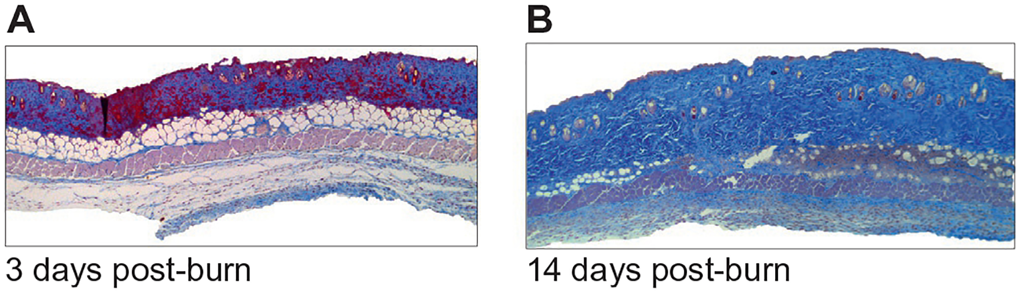
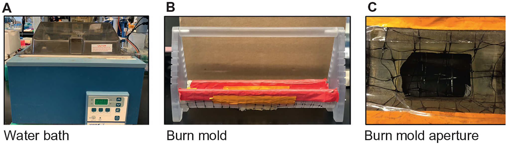
Burns are a severe form of trauma that account for 1.1 million cases necessitating medical attention and 4500 mortalities annually in the United States alone. Importantly, the initial trauma is succeeded by extensive, prolonged physiological alterations that detrimentally impact multiple organ systems. Given the complexity of post-burn pathophysiology, in vitro experiments are insufficient to model thermal injuries. Therefore, compatible animal burn models are essential for studying burn-related phenomena. In this chapter, we discuss commonly employed small animal burn models and their comparability and applicability to human studies. In particular, we compare post-burn wound healing between the species as well as relevant hypermetabolic and inflammatory characteristics, providing a better understanding of the pros and cons of utilizing a small animal surrogate for human burns. We further provide an overview of the rodent scald burn model methodology as well as a comparison between elderly, aged and young animals, providing a guide for tailoring animal model choice based on the relevant research question.
Keywords: Burn; Rodent model; Scald; Small animals.
Copyright © 2022 Elsevier Inc. All rights reserved.
Figures
Similar articles
-
Animal models in burn research.Cell Mol Life Sci. 2014 Sep;71(17):3241-55. doi: 10.1007/s00018-014-1612-5. Epub 2014 Apr 9. Cell Mol Life Sci. 2014. PMID: 24714880 Free PMC article. Review.
-
Large animal models of thermal injury.Methods Cell Biol. 2022;168:191-219. doi: 10.1016/bs.mcb.2021.12.015. Epub 2022 Jan 20. Methods Cell Biol. 2022. PMID: 35366983
-
Comparison of Thermal Burn-Induced and Excisional-Induced Scarring in Animal Models: A Review of the Literature.Adv Wound Care (New Rochelle). 2022 Mar;11(3):150-162. doi: 10.1089/wound.2021.0035. Epub 2021 Dec 10. Adv Wound Care (New Rochelle). 2022. PMID: 34841897 Review.
-
Enhanced wound-healing performance of a phyto-polysaccharide-enriched dressing - a preclinical small and large animal study.Int Wound J. 2017 Dec;14(6):1359-1369. doi: 10.1111/iwj.12813. Epub 2017 Sep 21. Int Wound J. 2017. PMID: 28941182 Free PMC article.
-
Mouse models in burns research: Characterisation of the hypermetabolic response to burn injury.Burns. 2020 May;46(3):663-674. doi: 10.1016/j.burns.2019.09.014. Epub 2019 Oct 10. Burns. 2020. PMID: 31606314
Cited by
-
Complex Hippocampal Response to Thermal Skin Injury and Protocols with Hyperbaric Oxygen Therapy and Filipendula ulmaria Extract in Rats.Int J Mol Sci. 2024 Mar 6;25(5):3033. doi: 10.3390/ijms25053033. Int J Mol Sci. 2024. PMID: 38474277 Free PMC article.
-
Therapeutic Effects of Nanochelating-Based Copper Nanoparticles on Burn Wound Healing in Mouse Model.Avicenna J Med Biotechnol. 2025 Jan-Mar;17(1):2-13. doi: 10.18502/ajmb.v17i1.17672. Avicenna J Med Biotechnol. 2025. PMID: 40094097 Free PMC article.
-
Lipolysis-derived linoleic acid drives beige fat progenitor cell proliferation.Dev Cell. 2022 Dec 5;57(23):2623-2637.e8. doi: 10.1016/j.devcel.2022.11.007. Dev Cell. 2022. PMID: 36473459 Free PMC article.
-
The efficacy of adipose-derived stem cells in burn injuries: a systematic review.Cell Mol Biol Lett. 2024 Jan 5;29(1):10. doi: 10.1186/s11658-023-00526-w. Cell Mol Biol Lett. 2024. PMID: 38182971 Free PMC article.
-
Mild burn amplifies the locomotive depression in demyelinated mice without muscle pathophysiological changes.PLoS One. 2024 Oct 7;19(10):e0308908. doi: 10.1371/journal.pone.0308908. eCollection 2024. PLoS One. 2024. PMID: 39374260 Free PMC article.
References
-
- Aggarwarl SJ, Shah SJ, Diller KR, & Baxter CR (1989). Fluorescence digital microscopy of interstitial macromolecular diffusion in burn injury. Computers in Biology and Medicine, 19(4), 245–261. - PubMed
-
- Ayala A, Herdon C, Lehman D, et al. (1995). The induction of accelerated thymic programmed cell death during polymicrobial sepsis: Control by corticosteroids not by tumor necrosis factor. Shock, 3, 259–267. - PubMed
Publication types
MeSH terms
Grants and funding
LinkOut - more resources
Full Text Sources
Miscellaneous
