. 2022 May;136(1):38-45.
doi: 10.1016/j.ymgme.2022.03.004. Epub 2022 Mar 18.

Comparative metabolomics in the Pahenu2 classical PKU mouse identifies cerebral energy pathway disruption and oxidative stress

Affiliations

Comparative metabolomics in the Pahenu2 classical PKU mouse identifies cerebral energy pathway disruption and oxidative stress

Steven F Dobrowolski et al. Mol Genet Metab. 2022 May.

Abstract

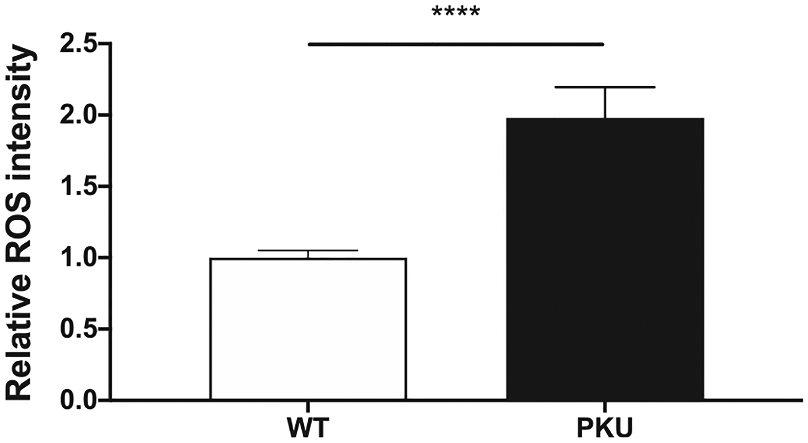
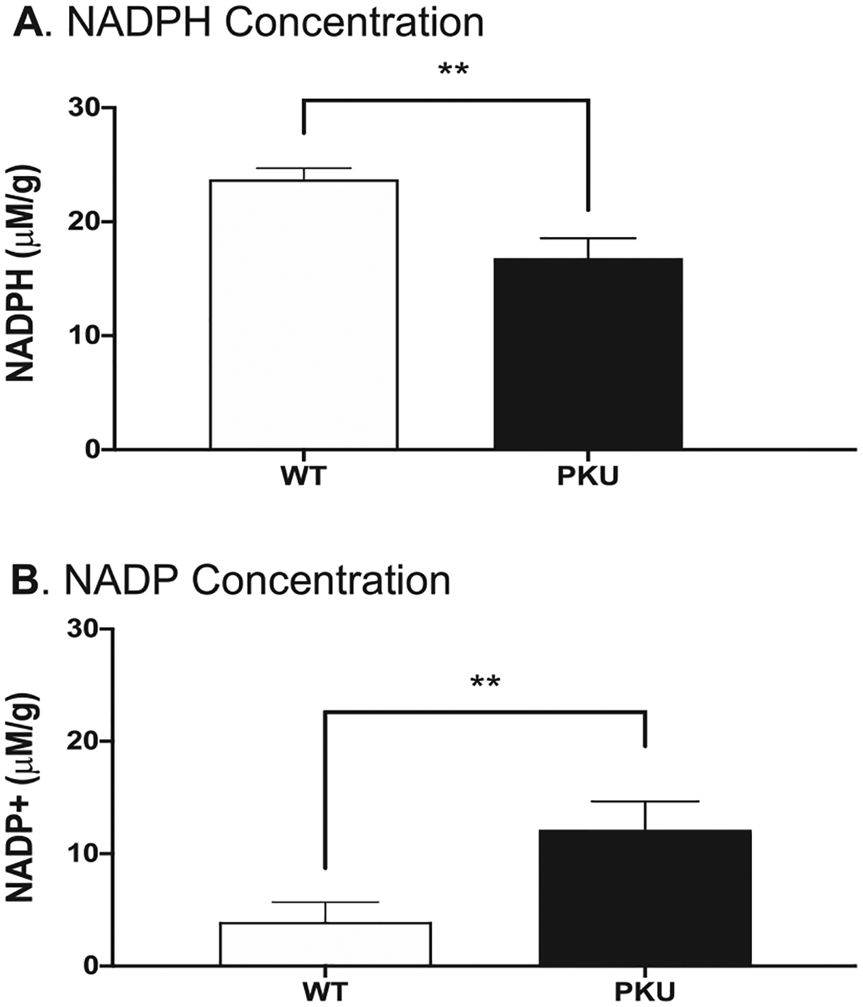
Classical phenylketonuria (PKU, OMIM 261600) owes to hepatic deficiency of phenylalanine hydroxylase (PAH) that enzymatically converts phenylalanine (Phe) to tyrosine (Tyr). PKU neurologic phenotypes include impaired brain development, decreased myelination, early onset mental retardation, seizures, and late-onset features (neuropsychiatric, Parkinsonism). Phe over-representation is systemic; however, tissue response to hyperphenylalaninemia is not consistent. To characterize hyperphenylalaninemia tissue response, metabolomics was applied to Pahenu2 classical PKU mouse blood, liver, and brain. In blood and liver over-represented analytes were principally Phe, Phe catabolites, and Phe-related analytes (Phe-conjugates, Phe-containing dipeptides). In addition to Phe and Phe-related analytes, the metabolomic profile of Pahenu2 brain tissue evidenced oxidative stress responses and energy dysregulation. Glutathione and homocarnosine anti-oxidative responses are apparent Pahenu2 brain. Oxidative stress in Pahenu2 brain was further evidenced by increased reactive oxygen species. Pahenu2 brain presents an increased NADH/NAD ratio suggesting respiratory chain complex 1 dysfunction. Respirometry in Pahenu2 brain mitochondria functionally confirmed reduced respiratory chain activity with an attenuated response to pyruvate substrate. Glycolysis pathway analytes are over-represented in Pahenu2 brain tissue. PKU pathologies owe to liver metabolic deficiency; yet, Pahenu2 liver tissue shows neither energy disruption nor anti-oxidative response. Unique aspects of metabolomic homeostasis in PKU brain tissue along with increased reactive oxygen species and respiratory chain deficit provide insight to neurologic disease mechanisms. While some elements of assumed, long standing PKU neuropathology are enforced by metabolomic data (e.g. reduced tryptophan and serotonin representation), energy dysregulation and tissue oxidative stress expand mechanisms underlying neuropathology.


Figures

Fig. 1.
Fig. 1.
A.Phe metabolism in Pahenu2blood, liver, and brain tissue. +analyte not observed in liver tissue; #analyte not observed in brain tissue B. Intrinsic cerebral anti-oxidative responses. Over-represented analytes are highlighted. Pathway analytes not identified by metabolomics or showing normal representation are in black font. Left side: Homocarnosine synthesis pathway. Chronic His over representation is demonstrated by conjugation reactions (similar to Phe conjugates see Fig. 1A) and the catabolite histamine. Right side: Glutathione pathway. ROS = reactive oxygen species C. Cerebral energy dysregulation. Over-represented analytes are highlighted. Pathway analytes not identified by metabolomics or showing normal representation are in black font. Upper Area: Pahenu2 frontal cortex over-represented glycolysis analytes. Lower Area: NADH over-representation with increased NADH/NAD ratio suggests respiratory chain complex 1 deficit, confirmed in Fig. 2 respirometry.
Fig. 1.
Fig. 1.
A.Phe metabolism in Pahenu2blood, liver, and brain tissue. +analyte not observed in liver tissue; #analyte not observed in brain tissue B. Intrinsic cerebral anti-oxidative responses. Over-represented analytes are highlighted. Pathway analytes not identified by metabolomics or showing normal representation are in black font. Left side: Homocarnosine synthesis pathway. Chronic His over representation is demonstrated by conjugation reactions (similar to Phe conjugates see Fig. 1A) and the catabolite histamine. Right side: Glutathione pathway. ROS = reactive oxygen species C. Cerebral energy dysregulation. Over-represented analytes are highlighted. Pathway analytes not identified by metabolomics or showing normal representation are in black font. Upper Area: Pahenu2 frontal cortex over-represented glycolysis analytes. Lower Area: NADH over-representation with increased NADH/NAD ratio suggests respiratory chain complex 1 deficit, confirmed in Fig. 2 respirometry.
Fig. 2.
Fig. 2.
Oxygen consumption rates by respirometry using fresh mouse frontal cortex mitochondria. Black arrows indicate substrate additions; Green font indicates flux response of Complex I (CI), Complex II (CII), and combined CI + CII. Mal = malate; ADP = adenosine diphosphate; Pyr = pyruvate; Glu = glutamate; Suc = succinate; Rot = rotenone
Fig. 3.
Fig. 3.
Reactive oxygen species in brain tissue. Brain tissue from six C57bl/6 control animals (3 male, 3 female) and six hyperphenylalaninemic Pahenu2 animals (3 male, 3 female) were assessed. **** p≤0.001
Fig. 4.
Fig. 4.
Frontal cortex tissue from six Pahenu2 and six C57bl/6 were assayed for NADP and NADPH. Fig. 4A. Quantification of NADPH. Fig. 4B. Quantification of NADP. ** p≤0.01 μM/g = micromole per gram tissue

Similar articles

Cited by

References

    1. Guthrie R, Sussi A, A simple phenylalanine method for detecting phenylketonuria in large populations of newborn infants, Pediatrics. 32 (1963) 318–322. - PubMed
    1. Bickel H, Gerrard J, Hickmans EM, Influence of phenylalanine intake on phenylketonuria, Lancet. 265 (1953) 812. - PubMed
    1. MacCready RA, Admissions of phenylketonuric patients to residential institutions before and after screening programs of the newborn infant, J. Pediatr 85 (1974) 383. - PubMed
    1. Azabdaftari A, van der Giet M, Schuchardt M, Hennermann JB, Plöckinger U, Querfeld U, The cardiovascular phenotype of adult patients with phenylketonuria, Orphanet J Rare Dis 14 (2019) 213. - PMC - PubMed
    1. Demirdas S, Coakley KE, Bisschop PH, Hollak CE, Bosch AM, Singh RH, Bone health in phenylketonuria: a systematic review and meta-analysis, Orphanet J Rare Dis 10 (2015) 17. - PMC - PubMed