. 2022 Jun;298(6):101882.
doi: 10.1016/j.jbc.2022.101882. Epub 2022 Mar 31.

Proteomic elucidation of the targets and primary functions of the picornavirus 2A protease

Affiliations

Proteomic elucidation of the targets and primary functions of the picornavirus 2A protease

Artem A Serganov et al. J Biol Chem. 2022 Jun.

Abstract

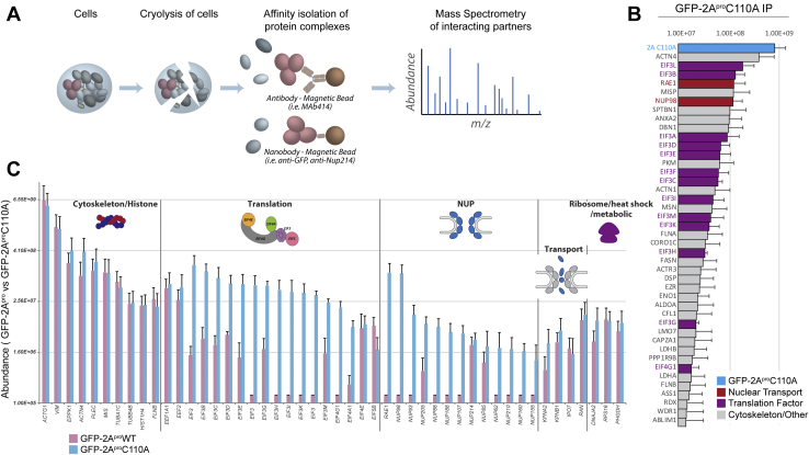
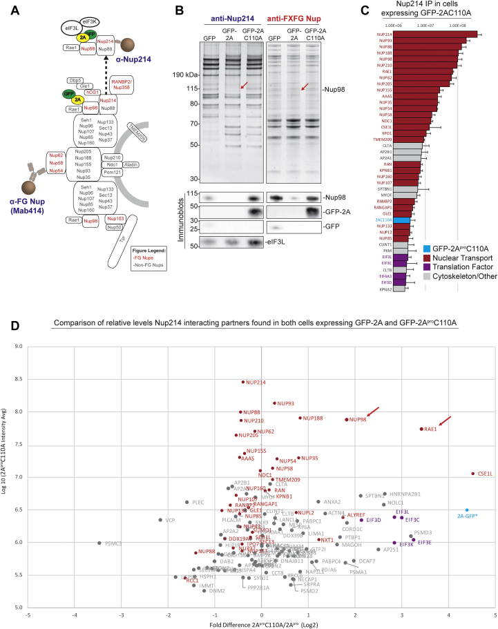
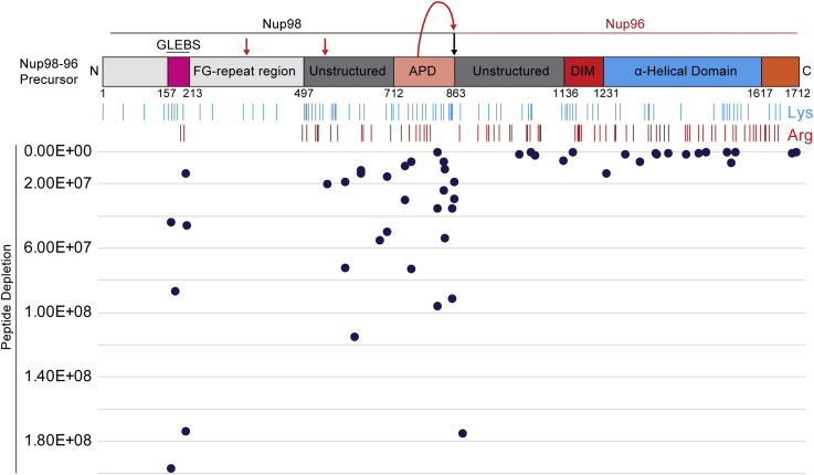
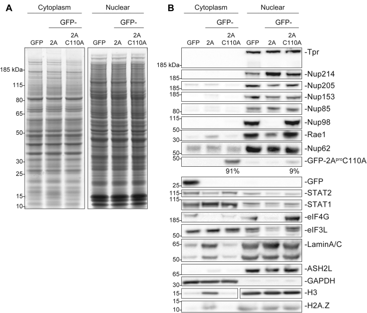
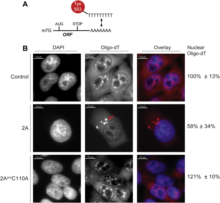
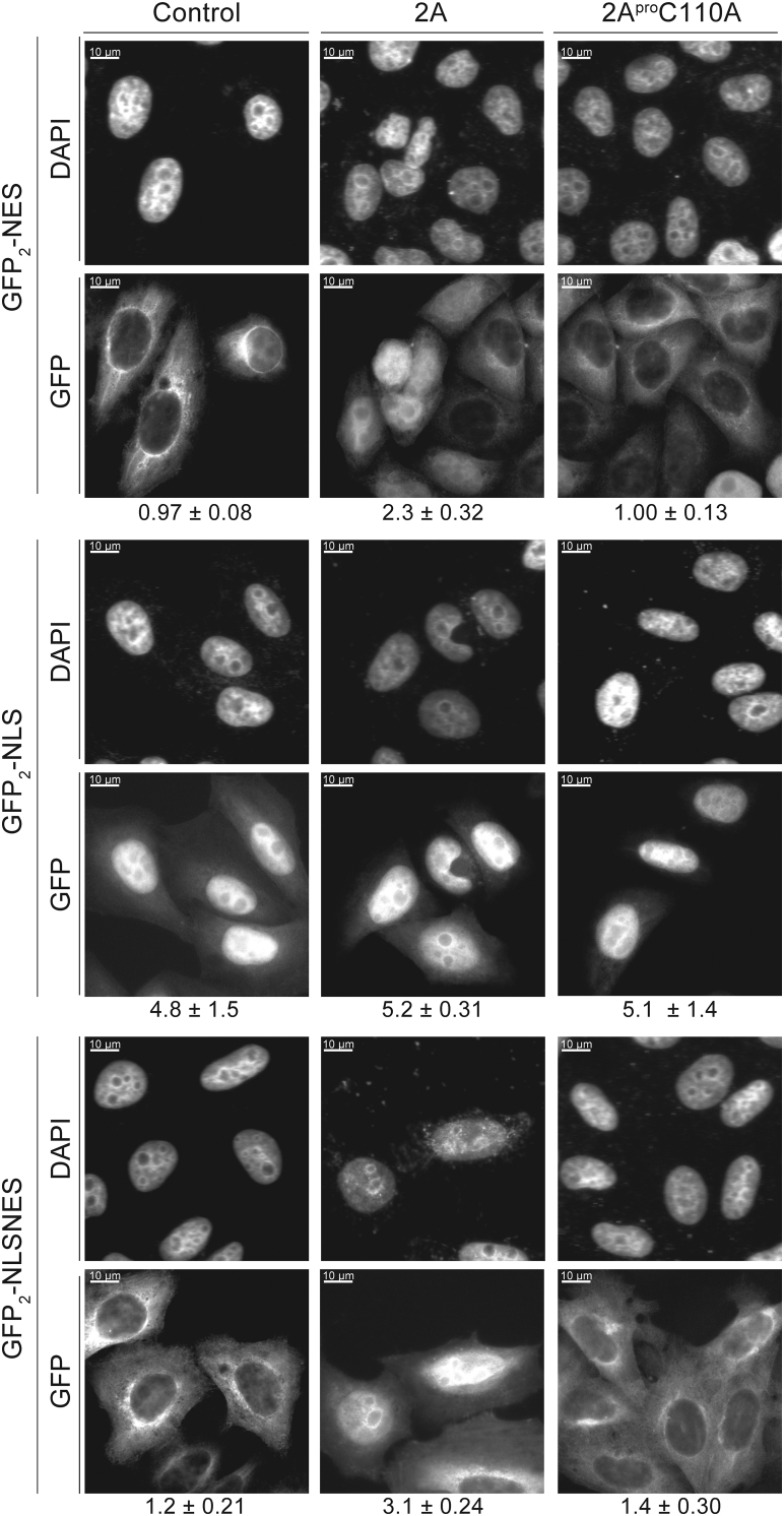
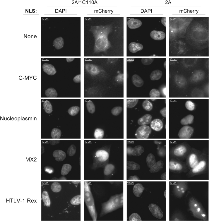
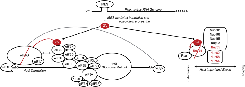
Picornaviruses are small RNA viruses that hijack host cell machinery to promote their replication. During infection, these viruses express two proteases, 2Apro and 3Cpro, which process viral proteins. They also subvert a number of host functions, including innate immune responses, host protein synthesis, and intracellular transport, by utilizing poorly understood mechanisms for rapidly and specifically targeting critical host proteins. Here, we used proteomic tools to characterize 2Apro interacting partners, functions, and targeting mechanisms. Our data indicate that, initially, 2Apro primarily targets just two cellular proteins: eukaryotic translation initiation factor eIF4G (a critical component of the protein synthesis machinery) and Nup98 (an essential component of the nuclear pore complex, responsible for nucleocytoplasmic transport). The protease appears to employ two different cleavage mechanisms; it likely interacts with eIF3L, utilizing the eIF3 complex to proteolytically access the eIF4G protein but also directly binds and degrades Nup98. This Nup98 cleavage results in only a marginal effect on nuclear import of proteins, while nuclear export of proteins and mRNAs were more strongly affected. Collectively, our data indicate that 2Apro selectively inhibits protein translation, key nuclear export pathways, and cellular mRNA localization early in infection to benefit viral replication at the expense of particular cell functions.

Keywords: mass spectrometry; nuclear pore; nuclear transport; picornavirus; plus-stranded RNA virus; protease; proteomics.


Conflict of interest statement

Conflict of interest The authors declare that they have no conflicts of interest with the contents of this article.

Figures

Figure 1
Figure 1
Biochemical isolation of 2Aprointeractome and binding specificity to Nup98. A, pipeline for affinity isolation of 2Apro interacting partners. B, GFP-2AproC110A interactome identified by MS (protein identity on Y-axis, peptide abundance on X-axis). C, side-by-side comparisons of protein interactomes of WT and mutant 2Apro affinity capture experiments. Protein identities are on the X-axis, organized by category of proteins (such as protein translation or Nup), with the Y-axis plotting peptide abundance. It is apparent that proteolytic activity causes a decrease in peptide abundance of a number of translation and transport related proteins. Adapted from Xiang Y. et al (2020).
Figure 2
Figure 2
Nup98 and eIF3 are primary interacting partners of the 2A protease during FG-Nup affinity capture. A, diagram of mammalian NPC with immunoprecipitation targets identified by specific antibody or nanobody (blue —Nup214, red—mAB414 specific for FG Nups). The protein complex (Nup214-Nup98-2AproC110A-eIF3L) proposed to be isolated during Nup214 affinity capture is shown above. B, affinity capture of Nup214, a cytoplasmic FG-Nup, and FXFG Nups reveals association with many NPC components. IP elutions were clarified by SDS-PAGE and analyzed by Silver stain (top panels) and immunoblotting (bottom panels). Nup214 and FG Nup IPs suggest 2Apro specifically cleaves Nup98. Immunoblot analysis suggest that immunopurifying GFP-2AproC110A, whether directly or indirectly (Nup214 IP) also correlates to an enrichment of eIF3L in eluates. C, top interacting partners identified by MS during Nup214 affinity capture (protein identity on Y-axis, peptide abundance on X-axis). D, affinity capture of Nup214 in the biological context of 2A and 2AproC110A expression reveals enrichment of Nup98, Rae1, and eIF3L, among other proteins, during 2AproC110A expression. Nup98 and Rae1 enrichment are due to cleavage of Nup98 in the 2Apro expressing cells and subsequent loss of Rae1 interaction to the Nup complex, while eIF3L and eIF3K are coprecipitating with 2AproC110A. The X-axis shows Log2 transformed fold-difference in label-free quantified MS abundance between datasets and the Y-axis measures 2AproC110A mean intensity.
Figure 3
Figure 3
Nup98 to 96 peptides plotted against secondary structure and assessed for depletion during 2Aproexpression. Nup98 is translated as a bicistronic fusion with 96 and separated into individual proteins by the autoproteolytic domain (APD). We plotted the label-free abundance of all Nup98 to 96 peptides identified by MS along the secondary structure of the fusion protein (X-axis) and observed depletion of Nup98 peptides originating only from its unstructured region specifically during 2Apro expression as measured and plotted along Y-axis (peptides located further down the Y-axis are more depleted). Red arrows indicate putative cleavage sites for 2Apro.
Figure 4
Figure 4
Subcellular fraction of nuclear proteins during 2Aproexpression. HeLa cells induced to express GFP, or catalytically active or inactive 2A protease, were subcellularly fractionated into cytoplasmic and nuclear fractions. A, Coomassie stain of fractions from fractionation steps, including the cytoplasmic and nuclear fractions. B, Western blot analysis of nuclear markers (H3, lamin A/C, ASH2L), a cytoplasmic marker (GAPDH), a variety of NPC structural (Tpr, Nup205, Nup85) and FG Nups (Nup214, Nup153, Nup98, Nup62), interferon-related pathways (STAT1, STAT2), as well as GFP, eIF3L, and Rae1. The depletion of specific Nups in the nuclear fraction in response to 2Apro expression is identified below the respective blot. Notably, Nup98 and Rae1 are significantly and specifically depleted due to 2Apro activity. Nup98 is degraded while Rae1 dissociates from the NPC due to lack of GLEBS motifs otherwise found on Nup98. GFP-2AproC110A can also be detected in the nuclear fraction as it associates through Nup98.
Figure 5
Figure 5
2Apro-mediated cleavage of Nup98 results in mRNA relocalization formation. A, Poly-T FISH was used to track mRNA export in cells. Tye563 is a red fluorescent dye that was conjugated to oligo-dT probes that bind the 3′ polyA tail of mRNA. B, 2Apro expressing cells were observed to have less mRNA signal and mislocalization of RNA speckles. This mislocalization was directly tied to the catalytic activity of the 2Apro, as the mutant protein did not affect mRNA signal. During 2Apro expression, RNA also seemed to localize to larger ‘speckles’ akin to P-bodies (red arrow). Signal reduction was assessed by measuring the total polyT FISH signal per cell and nuclei and comparing the average signal levels between each cell and across sample types.
Figure 6
Figure 6
GFP2-NLS/NES/NLSNES protein fusions are constitutively expressed in a HeLa stable cell line. Cells were either untreated (control—unperturbed signal) or transfected to express GFP-2Apro or GFP-2AproC110A. Cells were imaged under DAPI and FITC filters, with DAPI showcasing nuclear localization, and FITC demonstrating GFP2 reporter localization during various protease production contexts. We calculated the ratio of nuclear signal over cytoplasmic signal for GFP2 localization and observed a marked increase in nuclear localization of GFP2-NES during proteolytic activity. NES, nuclear export signal; NLS, nuclear localization signal.
Figure 7
Figure 7
2Apro-mediated cleavage of Nup98 results in localization changes for transport reporters with various NLS motifs. Fluorescent reporters consisting of an NLS motif and an mCherry tag, fused to a LacZ peptide. Cellular localization of NLS reporters was assayed with and without 2Apro expression. Nucleoplasmin, MX2, and C-Myc NLS reporters localized to the nucleus, even during 2Apro expression, suggesting those NLS motifs function via import pathways are not solely dependent on Nup98. The HTLV-1 Rex NLS motif also has RNA binding activity and shows mislocalization. HTLV-1, human T-cell leukemia virus type 1; NLS, nuclear localization signal; MX2, myxovirus resistance 2 protein.
Figure 8
Figure 8
Unified model of initial stages of picornaviral protease expression and function. Upon virion intake and genome release, the picornaviral RNA genome is translated as a polyprotein that is proteolytically processed into individual protein subunits by proteases 2A and 3C. The 2A protease proceeds to cleave Nup98 and eIF4G, with the latter cleavage facilitated by 2Apro association with the eIF3 complex. Cleavage of Nup98 dissociates Rae1 from the NPC, and the loss of both proteins from the NPC impacts nuclear transport of proteins and mRNA and relocalizes nuclear factors that promote internal ribosome entry site (IRES)-mediated translation. In parallel, eIF4G cleavage results in a shutdown of the host cell’s translation capability, diminishing the infected cells’ ability to react to the picornaviral infection. As the infection continues and additional viral proteins are translated, 2Apro activity results in dozens of secondary and tertiary cleavage targets, such as Matrin3, MAVS, and PABP, among many others (28). NPC, nuclear pore complex.

Similar articles

Cited by

References

    1. Fan Y., Sanyal S., Bruzzone R. Breaking bad: How viruses subvert the cell cycle. Front. Cell Microbiol. 2018;8:396. - PMC - PubMed
    1. Walsh D., Mohr I. Viral subversion of the host protein synthesis machinery. Nat. Rev. Microbiol. 2011;9:860–875. - PMC - PubMed
    1. Yarbrough M.L., Mata M.A., Sakthivel R., Fontoura B.M. Viral subversion of nucleocytoplasmic trafficking. Traffic. 2014;15:127–140. - PMC - PubMed
    1. Murphy P.M. Viral exploitation and subversion of the immune system through chemokine mimicry. Nat. Immunol. 2001;2:116–122. - PubMed
    1. Tang J.W., Holmes C.W. Acute and chronic disease caused by enteroviruses. Virulence. 2017;8:1062–1065. - PMC - PubMed

Publication types

MeSH terms

LinkOut - more resources