. 2022 May;298(5):101883.
doi: 10.1016/j.jbc.2022.101883. Epub 2022 Mar 31.

Mitochondria-associated myosin 19 processively transports mitochondria on actin tracks in living cells

Affiliations

Mitochondria-associated myosin 19 processively transports mitochondria on actin tracks in living cells

Osamu Sato et al. J Biol Chem. 2022 May.

Abstract

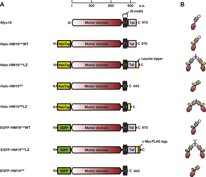
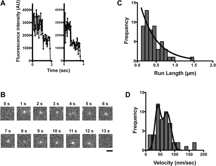
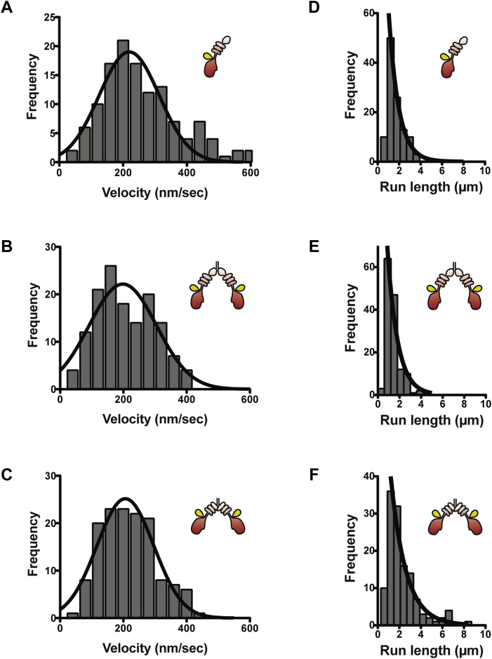
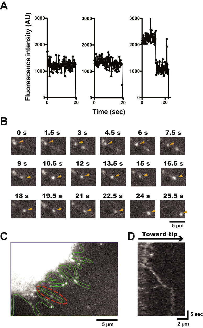
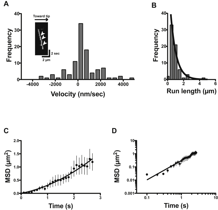
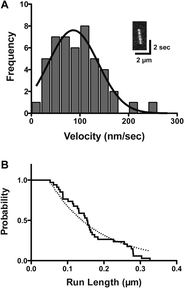
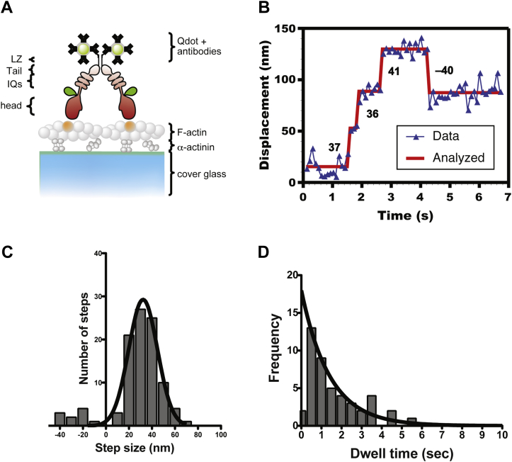
Mitochondria are fundamentally important in cell function, and their malfunction can cause the development of cancer, cardiovascular disease, and neuronal disorders. Myosin 19 (Myo19) shows discrete localization with mitochondria and is thought to play an important role in mitochondrial dynamics and function; however, the function of Myo19 in mitochondrial dynamics at the cellular and molecular levels is poorly understood. Critical missing information is whether Myo19 is a processive motor that is suitable for transportation of mitochondria. Here, we show for the first time that single Myo19 molecules processively move on actin filaments and can transport mitochondria in cells. We demonstrate that Myo19 dimers having a leucine zipper processively moved on cellular actin tracks in demembraned cells with a velocity of 50 to 60 nm/s and a run length of ∼0.4 μm, similar to the movement of isolated mitochondria from Myo19 dimer-transfected cells on actin tracks, suggesting that the Myo19 dimer can transport mitochondria. Furthermore, we show single molecules of Myo19 dimers processively moved on single actin filaments with a large step size of ∼34 nm. Importantly, WT Myo19 single molecules without the leucine zipper processively move in filopodia in living cells similar to Myo19 dimers, whereas deletion of the tail domain abolished such active movement. These results suggest that Myo19 can processively move on actin filaments when two Myo19 monomers form a dimer, presumably as a result of tail-tail association. In conclusion, Myo19 molecules can directly transport mitochondria on actin tracks within living cells.

Keywords: TIRF microscopy; intracellular movement; mitochondria; single molecule; unconventional myosin.


Conflict of interest statement

Conflict of interest The authors declare that they have no conflicts of interest with the contents of this article.

Figures

Figure 1
Figure 1
Schematic drawing of HM19 constructs used in this study.A, diagram of domain structures of HM19 constructs. HM19 consists of a motor domain (dark red), three IQ motifs (black), and a tail domain (pink). To observe single-molecule movement of HM19 in living cells, HaloTag was introduced at the N-terminal end (yellow green). To produce the dimer form of HM19 construct, a GCN4 leucine-zipper motif was introduced at the C-terminal end of HM19 (yellow). To purify the HM19 protein and perform the single-molecule stepping with Qdots, c-Myc/FLAG sequences were introduced at the C-terminal end of the HM19 constructs (orange). To confirm the localization of HM19 construct, and single molecular experiments both in cells and in vitro, EGFP-HM19 was also made (green). The number at the top of the panel represents amino acid residues of Myo19. B, configuration of HM19 constructs. It is speculated that HM19 dimer is formed when a GCN4 leucine-zipper motif is attached to the C-terminal end of HM19 construct. EGFP, enhanced GFP; HM19, human Myo19; Qdot, quantum dot.
Figure 2
Figure 2
Movement of HM19FullLZ-associated mitochondria vesicles on demembraned cells. The movement of Halo-HM19FullLZ-associated HMF was observed with a TIRF microscope at 5 fps in the presence of 1 mM ATP, and the run length and velocity were analyzed. A, representative movement of HMF prepared from HM19FullLZ-expressing cells on demembraned U2OS cells. Halo-HM19FullLZ was expressed in HEK293T cells and stained with R110 direct reagent. The HMF prepared as described in the “Experimental procedures” section was added to demembraned U2OS cells, and the movement was observed with a TIRF microscope in the presence of 1 mM ATP (Movie S1). The bar represents 1 μm. B, run length of HM19FullLZ-associated mitochondria vesicles. The position of the moving light spot was tracked, and the run length was calculated. The solid line shows the best fit to a single exponential equation, R0er/λ, where R0 is the initial frequency extrapolated to zero run length, r is the run length, and λ is the average run length. The average run length was 0.37 ± 0.09 μm (SEM, n = 48). C, the velocity of HM19FullLZ-associated mitochondria vesicles. The distribution was fitted to two Gaussian equations with the mean velocity of 27.8 ± 1.0 and 61.9 ± 6.4 nm/s (SEM, n = 48), respectively. HEK293T, human embryonic kidney 293T cell line; HM19, human Myo19; HMF, heavy mitochondrial fraction; LZ, leucine zipper; R110, rhodamine 110; TIRF, total internal reflection fluorescence.
Figure 3
Figure 3
Single-molecule imaging of purified HM19FullLZ on demembraned cells. EGFP-HM19FullLZ was purified, and the movement on demembraned U2OS cells was observed with a TIRF microscope at 5 fps. A, representative two-step photobleaching of the fluorescent spots of EGFP-HM19FullLZ. B, typical time-lapse images of EGFP-HM19FullLZ movement in the presence of 1 mM ATP. EGFP-HM19FullLZ single-molecule movement from 0.2 to 12.6 s was recorded. The bar represents 1 μm. The pixel-by-pixel size is 94 nm. C, run length of EGFP-HM19FullLZ in the presence of 1 mM ATP. The position of moving light spot was tracked, and the run length was calculated. Solid line shows the best fit to a single exponential equation, R0er/λ. The average run length was 0.43 ± 0.08 μm (SEM, n = 55). D, the velocity of EGFP-HM19FullLZ in the presence of 1 mM ATP. The distribution was fitted to two Gaussian equations with the mean velocities of 41.1 ± 6.4 and 73.3 ± 8.6 nm/s (SEM), respectively, for EGFP-HM19FullLZ. AU, arbitrary unit; EGFP, enhanced GFP; HM19, human Myo19; LZ, leucine zipper; TIRF, total internal reflection fluorescence.
Figure 4
Figure 4
Movement of HM19 constructs in living cells. Cultured HeLa cells were transfected with Halo-HM19 constructs and stained with R110 Direct HaloTag ligands according to the “Experimental procedures” section. The movement in filopodia was then observed at 1 or 2 fps with a TIRF microscope by HILO illumination. The run length and velocities were determined as described in the “Experimental procedures” section. AC, the histograms of velocities of Halo-HM19 constructs in filopodia. (A) R110-Halo-HM19FullWT, (B) R110-Halo-HM19FullLZ, and (C) R110-Halo-HM19IQ3LZ. The mean velocities were 0.263 ± 0.012 μm/s (SEM, n = 127) for R110-Halo-HM19FullWT, 0.209 ± 0.008 μm/s (SEM, n = 140) for R110-Halo-HM19FullLZ and 0.219 ± 0.008 μm/s (SEM, n = 128) for R110-Halo-HM19IQ3LZ. The distributions were fitted to a Gaussian equation (solid lines). The movement toward filopodial tip was analyzed. DF, the histograms of run lengths of R110-Halo-HM19s in filopodia. (D) R110-Halo-HM19FullWT, (E) R110-Halo-HM19FullLZ, and (F) R110-Halo-HM19IQ3LZ. The solid line shows the best fit to a single exponential equation, R0er/λ. The calculated average run lengths for R110-Halo-HM19FullWT, R110-Halo-HM19FullLZ, and R110-Halo-HM19IQ3LZ were 0.91 ± 0.08 μm (SEM, n = 127), 0.95 ± 0.17 μm (SEM, n = 140), and 1.4 ± 0.2 μm (SEM, n = 128), respectively. The movement toward filopodial tip was analyzed. HILO, highly inclined and laminated optical sheet; HM19, human Myo19; LZ, leucine zipper; R110, rhodamine 110; TIRF, total internal reflection fluorescence.
Figure 5
Figure 5
Single-molecule imaging of HM19FullWT in living cells. Cultured HeLa cells were transfected with Halo-HM19FullWT and stained with R110 Direct HaloTag ligands, and the movement of R110-Halo-HM19FullWT molecules in filopodia was observed with a TIRF microscope by HILO illumination. A, representative photobleaching of the fluorescent spots of R110-Halo-HM19FullWT. Most of the fluorescent spots show one-step photobleaching, but two-step photobleaching can also be found. The images were captured at 2 fps. B, time-lapse images of R110-Halo-HM19FullWT movement in living HeLa cells. Moving fluorescent spots are indicated by arrowheads. The images were captured at 2 fps. C, representative image of living HeLa cells. The profile of filopodia was shown in green lines. The broken red line shows a typical filopodia used for the kymograph analysis shown in (D). Kymograph shows the fluorescent spot continuously move in filopodia. HILO, highly inclined and laminated optical sheet; HM19, human Myo19; R110, rhodamine 110; TIRF, total internal reflection fluorescence.
Figure 6
Figure 6
Diffusive component and mean square displacement (MSD) analysis of the movement of HM19FullWT in living cells.A, velocity distribution of R110-Halo-HM19FullWT movement. R110-Halo-HM19FullWT movements in living HeLa cells were captured at fast frame rate (10 fps), and the velocities were determined from the kymographs (n = 106). The inset in (A) shows a typical kymograph of R110-Halo-HM19FullWT movement. By monitoring the movement with fast frame rate, occasional jumps were observed along with continuous directional movement (arrowheads). These jumps, presumably because of diffusion, were observed in both plus and minus directions. The forward and backward movements within filopodia are shown in the figure. It is expected that these fast diffusive components of plus and minus directions can be canceled out when the movement is monitored with slow frame rate. B, run length distribution of R110-Halo-HM19FullWT movement. Solid line shows the best fit to a single exponential equation, R0e−r/λ. The calculated average run length for R110-Halo-HM19FullWT was 0.70 ± 0.08 μm (SEM, n = 67). The movement toward filopodial tip was analyzed. C, MSD analysis for R110-Halo-HM19FullWT movement. The position of moving light spot was tracked using 2D fitting and tracking software, and the MSD was calculated. The plot is fitted with an equation of f(t) = 4Dt + v2t2 for 0 to 2.7 s (solid line, v = 0.33 ± 0.07 μm/s, D = 0.05 ± 0.02 μm2/s), where v = velocity; t = time; and D = diffusion coefficient. Error bars represent SEM (n = 9). D, log–log plot of MSD analysis for R110-Halo-HM19FullWT. The same data in (C) were plotted in the log–log axes and fitted with f(t) = ctα, where c = constant, t = time, and α = slope of the plot (solid line). The α value of the plot for R110-Halo-HM19FullWT is calculated to be 1.47 ± 0.08. Error bars represent SEM. HM19, human Myo19; R110, rhodamine 110.
Figure 7
Figure 7
Velocity and run length of HM19FullLZ on single actin filaments in vitro. The single-molecule movement of purified EGFP-HM19FullLZ on single actin filaments was observed with a TIRF microscope. Experiments were carried out in the presence of 1 mM ATP using glass flow chambers in which actin filaments were immobilized (see the “Experimental procedures” section), and the fluorescence images were captured at 10 fps. A, the velocity of EGFP-HM19FullLZ on single actin filaments. The mean velocity was 95.1 ± 7.4 nm/s (SEM, n = 47). The solid line shows the best fit to a Gaussian equation. Inset shows a typical kymograph of the EGFP-HM19FullLZ movement. B, run length of EGFP-HM19FullLZ on single actin filaments. The graph was shown with a Kaplan–Meier survival curve. Broken line shows the best fit to the single exponential equation as described for Figure 3. The average run length was 0.18 ± 0.01 μm (SEM, n = 47). EGFP, enhanced GFP; HM19, human Myo19; LZ, leucine zipper; TIRF, total internal reflection fluorescence.
Figure 8
Figure 8
Step-size and dwell time distribution of HM19FullLZ-Qdot on single actin filaments. The movement of EGFP-HM19FullLZ-Qdot525 (see the “Experimental procedures” section) on single actin filaments was observed with a TIRF microscope. A, schematic drawing of EGFP-HM19FullLZ-Qdot525. The Qdots were attached to the C-terminal c-Myc tag of EGFP-HM19FullLZ through first (anti-c-Myc) and second (antimouse Fab’) antibodies. B, a representative trace of EGFP-HM19FullLZ-Qdot525 stepping. The experiment was carried out in the presence of 2 μM ATP, and the fluorescence images were captured at 10 fps. Solid line shows the best fit to the trajectory. The number in the panel is the displacement of each step in nanometer. C, step size distribution of EGFP-HM19FullLZ-Qdot525. The mean step size of forward and backward steps is 33.8 ± 1.3 nm (mean ± SEM, n = 91) and −27.2 ± 3.3 nm (SEM, n = 10), respectively. Black solid line shows the best fit to a Gaussian equation. D, dwell time distribution of EGFP-HM19FullLZ-Qdot525 in the presence of 2 μM ATP. Solid line shows the best fit to a single exponential equation, kekt, where t is time, and k is rate constant (the first bin is excluded from the fitting). The average waiting time (τ = 1/k) is 1.4 ± 0.1 s (SEM, n = 46). EGFP, enhanced GFP; HM19, human Myo19; LZ, leucine zipper; Qdot, quantum dot; TIRF, total internal reflection fluorescence.

Similar articles

Cited by

References

    1. Coluccio L.M. Springer; Dordrecht: 2008. Myosins: A Superfamily of Molecular Motors.
    1. Odronitz F., Kollmar M. Drawing the tree of eukaryotic life based on the analysis of 2,269 manually annotated myosins from 328 species. Genome Biol. 2007;8:R196. - PMC - PubMed
    1. Yu C., Feng W., Wei Z., Miyanoiri Y., Wen W., Zhao Y., Zhang M. Myosin VI undergoes cargo-mediated dimerization. Cell. 2009;138:537–548. - PubMed
    1. Umeki N., Jung H.S., Sakai T., Sato O., Ikebe R., Ikebe M. Phospholipid-dependent regulation of the motor activity of myosin X. Nat. Struct. Mol. Biol. 2011;18:783–788. - PubMed
    1. Sakai T., Umeki N., Ikebe R., Ikebe M. Cargo binding activates myosin VIIA motor function in cells. Proc. Natl. Acad. Sci. U. S. A. 2011;108:7028–7033. - PMC - PubMed

Publication types

LinkOut - more resources