. 2022 Jul 15:208:114200.
doi: 10.1016/j.bios.2022.114200. Epub 2022 Mar 22.

Probing the mutation independent interaction of DNA probes with SARS-CoV-2 variants through a combination of surface-enhanced Raman scattering and machine learning

Affiliations

Probing the mutation independent interaction of DNA probes with SARS-CoV-2 variants through a combination of surface-enhanced Raman scattering and machine learning

Parikshit Moitra et al. Biosens Bioelectron. .

Abstract

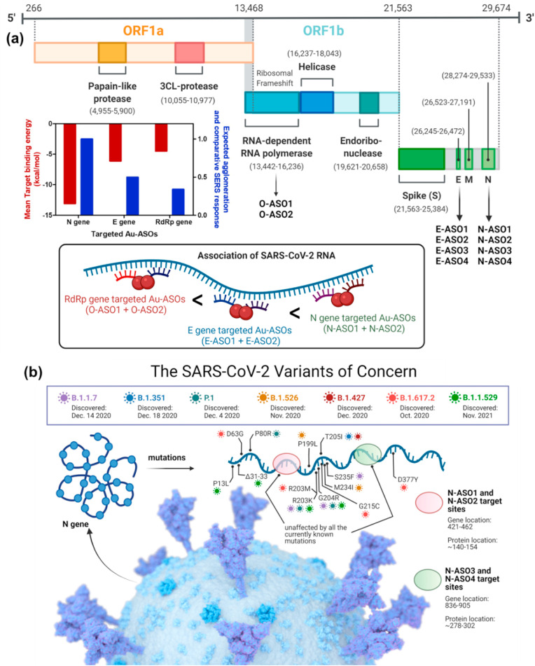
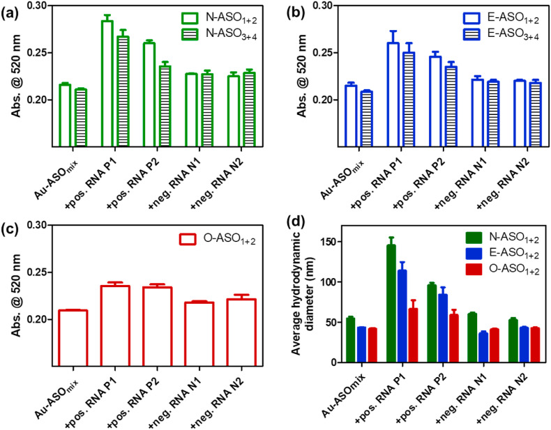
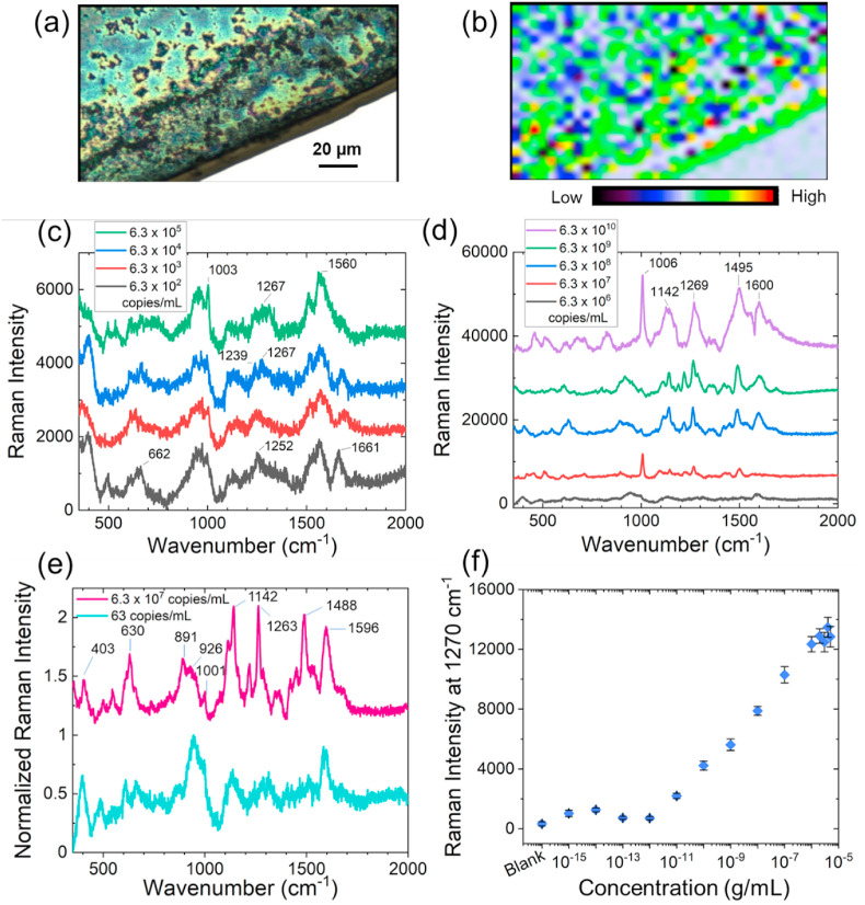
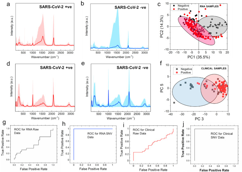
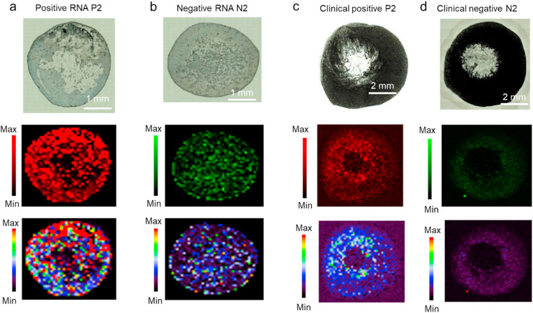
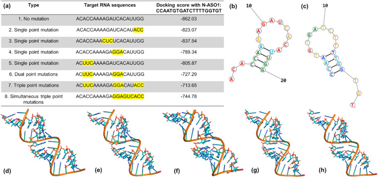
Severe acute respiratory syndrome coronavirus 2 (SARS-CoV-2) evolution has been characterized by the emergence of sets of mutations impacting the virus characteristics, such as transmissibility and antigenicity, presumably in response to the changing immune profile of the human population. The presence of mutations in the SARS-CoV-2 virus can potentially impact therapeutic and diagnostic test performances. We design and develop here a unique set of DNA probes i.e., antisense oligonucleotides (ASOs) which can interact with genetic sequences of the virus irrespective of its ongoing mutations. The probes, developed herein, target a specific segment of the nucleocapsid phosphoprotein (N) gene of SARS-CoV-2 with high binding efficiency which do not mutate among the known variants. Further probing into the interaction profile of the ASOs reveals that the ASO-RNA hybridization remains unaltered even for a hypothetical single point mutation at the target RNA site and diminished only in case of the hypothetical double or triple point mutations. The mechanism of interaction among the ASOs and SARS-CoV-2 RNA is then explored with a combination of surface-enhanced Raman scattering (SERS) and machine learning techniques. It has been observed that the technique, described herein, could efficiently discriminate between clinically positive and negative samples with ∼100% sensitivity and ∼90% specificity up to 63 copies/mL of SARS-CoV-2 RNA concentration. Thus, this study establishes N gene targeted ASOs as the fundamental machinery to efficiently detect all the current SARS-CoV-2 variants regardless of their mutations.

Keywords: Antisense oligonucleotide; Mutation resistant probe; SARS-CoV-2 variants; Selective and ultrasensitive diagnosis; Surface-enhanced Raman scattering.


Conflict of interest statement

The authors declare that they have no known competing financial interests or personal relationships that could have appeared to influence the work reported in this paper.

D.P. is the founder or co-founder for four University based start ups. None of these entities, however, supported this work.

Figures

Fig. 1
Fig. 1
(a) Genomic organization of SARS-CoV-2 and its schematic agglomeration pattern with gold nanoparticles differentially functionalized with antisense oligonucleotides targeted towards different genetic segments; (b) Currently known N gene mutation sites for SARS-CoV-2 and their alignment with the targeted sites of the developed ASOs.
Fig. 2
Fig. 2
Aggregation induced change in absorbance at 520 nm for the differentially functionalized Au-ASO NPs targeted for (a) N gene; (b) E gene and (c) RdRp gene upon the addition of RNAs (1 μL) extracted from clinically positive and negative SARS-CoV-2 samples. (d) Comparative change in average hydrodynamic diameter among the different Au-ASO NPs upon the addition of RNAs. In each case, the mixture was incubated for 15 min at room temperature prior to recording the change in absorbance. Ct value of positive RNA P1 and P2 are 14.7 and 27 respectively by RT-PCR analyses which correspond to 107186 and 781 copies/μL of SARS-CoV-2 RNA.
Fig. 3
Fig. 3
(a) Bright field image of N-ASO1+2 capped AuNPs admixed with SARS-CoV-2 RNA; (b) Raman microscopic image of SARS-CoV-2 RNA associated N-ASO1+2 capped AuNPs in the range of 1495–1602 cm-1 (785 nm laser, 100% power, grating of 1200, 50 XL magnification) with the center of 1200 cm-1. Red means higher concentration and blue indicates lower concentration of sample; SERS spectra of N-ASO1+2 capped AuNPs with (c) lower, and (d) higher SARS-CoV-2 RNA concentrations; (e) Comparison of spectral figures at low and high SARS-CoV-2 RNA concentrations from the normalized Raman spectra of N-ASO1+2 AuNPs. The spectral positions and shapes are found to be similar at low and high concentrations; (f) Limit of detection of N-ASO1+2 capped AuNPs towards the detection of SARS-CoV-2 RNA. Here, 1 fg/mL corresponds to 63 copies/mL of SARS-CoV-2 RNA.
Fig. 4
Fig. 4
Representative Raman spectra of N-ASO1+2 capped AuNPs added with RNA extracted from SARS-CoV-2 (a) positive, P1 and (b) negative, N1 clinical samples; The respective representative Raman spectra of N-ASO1+2 capped AuNPs admixed with direct SARS-CoV-2 (d) positive and (e) negative clinical samples are also shown without the extraction of RNA but with just the addition of lysis buffer. The range of all the spectra is shown as shaded color for each sample while the dark color is representing the mean spectra. PCA score plots show the separation between negative and positive samples of (c) extracted RNA samples and (f) direct clinical samples; ROC curves for (g) raw data and (h) after performing SNV operation on the SERS data obtained with extracted RNA samples; ROC curves for (i) raw data and (j) after performing SNV operation on the SERS data obtained while analyzing the direct clinical samples without any extraction of RNA from N-ASO1+2 capped AuNPs.
Fig. 5
Fig. 5
Representative Raman microscopic images of N-ASO1+2 capped AuNPs admixed with SARS-CoV-2 positive and negative (a, b) extracted RNA and (c, d) direct clinical samples at 928 cm-1 using StreamHR acquisition mode and obtained with 785 nm laser, 100% power, grating of 1200, magnification of 50 XL and exposure time of 0.5 s. The pixel size for RNA and clinical samples were 70 μm × 70 μm and 200 μm × 200 μm, respectively. Distribution for whole Raman spectrum is from 200 to 3200 cm-1. For RNA samples, map has around 1500 pixels (that is 1500 Raman spectra) and for clinical samples, map has around 2500 pixels (that is 2500 Raman spectra).
Fig. 6
Fig. 6
(a) List of hypothetical single, double, and triple point mutations on the RNA site targeted by N-ASO1. The hypothetical mutation sites are highlighted with yellow background; (b) Theoretical model of RNA with no mutation; (c) Theoretical model of N-ASO1; Docked model of N-ASO1 with RNA having (d) no mutation sites; (e) single point mutation as shown in no. 4; (f) double point mutations as shown in no. 6; (g) triple point mutations as shown in no. 7 and (h) simultaneous triple point mutations as shown in no. 8.

References

    1. Abid Hasan S.M., He Y., Chang T.-W., Wang J., Gartia M.R. Detecting DNA methylation using surface-enhanced Raman spectroscopy. J. Phys. Chem. C. 2019;123:698–709. doi: 10.1021/acs.jpcc.8b10178. - DOI
    1. Afzal A. Molecular diagnostic technologies for COVID-19: limitations and challenges. J. Adv. Res. 2020;26:149–159. doi: 10.1016/j.jare.2020.08.002. - DOI - PMC - PubMed
    1. Alafeef M., Dighe K., Moitra P., Pan D. Rapid, ultrasensitive, and quantitative detection of SARS-CoV-2 using antisense oligonucleotides directed electrochemical biosensor chip. ACS Nano. 2020;14:17028–17045. doi: 10.1021/acsnano.0c06392. - DOI - PMC - PubMed
    1. Alafeef M., Moitra P., Dighe K., Pan D. Hyperspectral mapping for the detection of SARS-CoV-2 using nanomolecular probes with Yoctomole sensitivity. ACS Nano. 2021;15:13742–13758. doi: 10.1021/acsnano.1c05226. - DOI - PMC - PubMed
    1. Alafeef M., Moitra P., Dighe K., Pan D. RNA-extraction-free nano-amplified colorimetric test for point-of-care clinical diagnosis of COVID-19. Nat. Protoc. 2021;16:3141–3162. doi: 10.1038/s41596-021-00546-w. - DOI - PubMed

Supplementary concepts