

Action of Hyaluronic Acid as a Damage-Associated Molecular Pattern Molecule and Its Function on the Treatment of Temporomandibular Disorders
- PMID: 35369538
- PMCID: PMC8971669
- DOI: 10.3389/fpain.2022.852249
Action of Hyaluronic Acid as a Damage-Associated Molecular Pattern Molecule and Its Function on the Treatment of Temporomandibular Disorders
Abstract
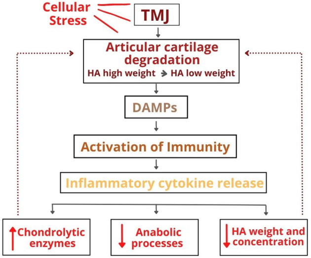
The temporomandibular joint is responsible for fundamental functions. However, mechanical overload or microtraumas can cause temporomandibular disorders (TMD). In addition to external factors, it is known that these conditions are involved in complex biological mechanisms, such as activation of the immune system, activation of the inflammatory process, and degradation of extracellular matrix (ECM) components. The ECM is a non-cellular three-dimensional macromolecular network; its most studied components is hyaluronic acid (HA). HA is naturally found in many tissues, and most of it has a high molecular weight. HA has attributed an essential role in the viscoelastic properties of the synovial fluid and other tissues. Additionally, it has been shown that HA molecules can contribute to other mechanisms in the processes of injury and healing. It has been speculated that the degradation product of high molecular weight HA in healthy tissues during injury, a low molecular weight HA, may act as damage-associated molecular patterns (DAMPs). DAMPs are multifunctional and structurally diverse molecules that play critical intracellular roles in the absence of injury or infection. However, after cellular damage or stress, these molecules promote the activation of the immune response. Fragments from the degradation of HA can also act as immune response activators. Low molecular weight HA would have the ability to act as a pro-inflammatory marker, promoting the activation and maturation of dendritic cells, the release of pro-inflammatory cytokines such as interleukin 1 beta (IL-1β), and tumor necrosis factor α (TNF-α). It also increases the expression of chemokines and cell proliferation. Many of the pro-inflammatory effects of low molecular weight HA are attributed to its interactions with the activation of toll-like receptors (TLRs 2 and 4). In contrast, the high molecular weight HA found in healthy tissues would act as an anti-inflammatory, inhibiting cell growth and differentiation, decreasing the production of inflammatory cytokines, and reducing phagocytosis by macrophages. These anti-inflammatory effects are mainly attributed to the interaction of high-weight HA with the CD44 receptor. In this study, we review the action of the HA as a DAMP and its functions on pain control, more specifically in orofacial origin (e.g., TMD).
Keywords: damage-associated molecular patterns; extracellular matrix; hyaluronic acid; synovial fluid; temporomadibular joint disorders.
Copyright © 2022 Ferreira, Sanz, Raybolt, Pereira and DosSantos.
Conflict of interest statement
The authors declare that the research was conducted in the absence of any commercial or financial relationships that could be construed as a potential conflict of interest.
Figures
References
-
- Okeson JF. Management of Temporomandibular Disorders and Occlusion. St. Louis, MI: Elsevier; (2008).
Publication types
LinkOut - more resources
Full Text Sources
Miscellaneous