Case Reports
. 2022 Mar 27;17(5):1798-1802.
doi: 10.1016/j.radcr.2022.03.006. eCollection 2022 May.

Pigmented villonodular synovitis of the knee in a child: a case report

Affiliations
Case Reports

Pigmented villonodular synovitis of the knee in a child: a case report

Dang Hong Hoa et al. Radiol Case Rep. .

Abstract

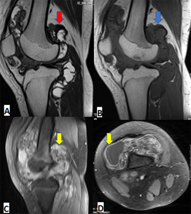
Pigmented villonodular synovitis is a rare proliferative process, especially in children. Pigmented villonodular synovitis can affect the synovial joint, tendon sheaths, and bursa membranes. Within synovial joint involvement, it is commonly seen in the knee joint but hip, ankle, shoulder, wrist, and other joints can be involved. The appearance characteristic is found on a magnetic resonance imaging scan. Complete excision and synovectomy are the usual treatment. In this article, we report a case of pigmented villonodular synovitis of the knee in a 12- year-old girl who underwent total synovectomy after the diagnosis was confirmed by biopsy. Three years after surgery, neither recurrence nor joint degeneration was found. The osteochondral defect at the tibial plateau was filled with calcium phosphate bone paste.

Keywords: Knee; Pigmented villonodular, Child; Synovitis.


Figures

Fig 1
Fig. 1
Ultrasound. Joint effusion (green arrow), with hypoechoic and irregular thickening of the synovium (red arrow).
Fig 2
Fig. 2
Computed tomography. Joint effusion (red arrow), diffuse irregular thickening of the synovium (yellow arrow), associated with the formation of subchondral bone cysts (blue arrow).
Fig 3
Fig. 3
Magnetic resonance imaging. Diffuse irregular thickening of the synovium in the knee joint, slightly hypointense in T1W (blue arrow), hypointense in T2W due to hemosiderin deposition (red arrow), after contrast the synovium show strong enhancement (yellow arrow).
Fig 4
Fig. 4
Knee arthroscopy: The knee synovial membrane is thickened, villonodular synovial hyperplasia throughout the posterior patellar space and the knee joint space. Nodular proliferation (green arrow), villous proliferation (red arrow).
Fig 5
Fig. 5
Histopathology. Microscopic sections show mononuclear cells admixed with osteoclast – like multinucleated giant cells and inflammatory cells (HE A – 40x, B-100x).

References

    1. Sullivan CJ, Eustace SJ, Kavanagh EC. Pigmented villonodular synovitis of the hip joint: three cases demonstrating characteristic MRI features. Radiol Case Rep. 24 Jun 2020;15(8):1335–1338. doi: 10.1016/j.radcr.2020.05.067. PMID: 32617126; PMCID: PMC7322487. - DOI - PMC - PubMed
    1. Fecek C, Carter KR. StatPearls [Internet] StatPearls Publishing; Treasure Island (FL): 7 Nov 2021. Pigmented villonodular synovitis. 2022 Jan–. PMID: 31751040. - PubMed
    1. Tritschler P, Baudrez V, Mutijima E. Diffuse pigmented villonodular synovitis of the subtalar joint. J Belg Soc Radiol. 31 Jan 2018;102(1):11. doi: 10.5334/jbsr.1477. PMID: 30039025; PMCID: PMC6032406. - DOI - PMC - PubMed
    1. Fałek A, Niemunis-Sawicka J, Wrona K, Szczypiór G, Rzepecka-Wejs L, Cięszczyk K, et al. Pigmented villonodular synovitis. Folia Med Cracov. 2018;58(4):93–104. PMID: 30745604. - PubMed
    1. Bernthal NM, Ishmael CR, Burke ZDC. Management of Pigmented villonodular synovitis (PVNS): an orthopedic surgeon's perspective. Curr Oncol Rep. 4 Jun 2020;22(6):63. doi: 10.1007/s11912-020-00926-7. PMID: 32494892. - DOI - PubMed

Publication types

LinkOut - more resources