Case Reports
. 2022 Mar 27;17(5):1803-1809.
doi: 10.1016/j.radcr.2022.03.014. eCollection 2022 May.

Lifesaving diagnosis of placenta accreta spectrum using MRI: Report of five cases

Affiliations
Case Reports

Lifesaving diagnosis of placenta accreta spectrum using MRI: Report of five cases

Belinda Koesmarsono et al. Radiol Case Rep. .

Abstract

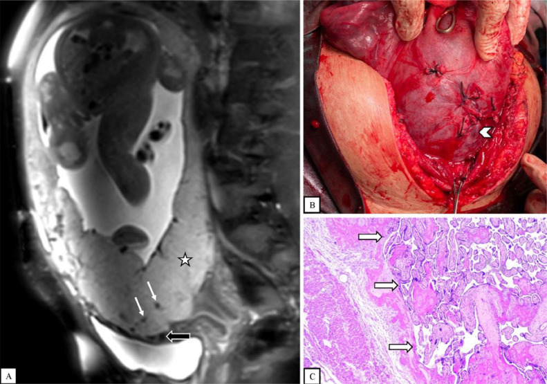
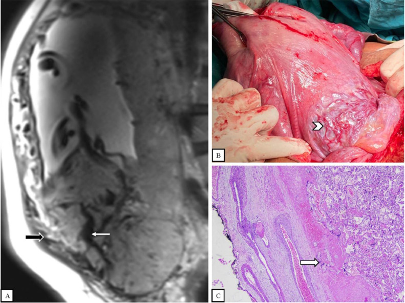
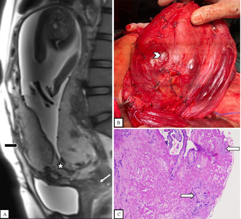
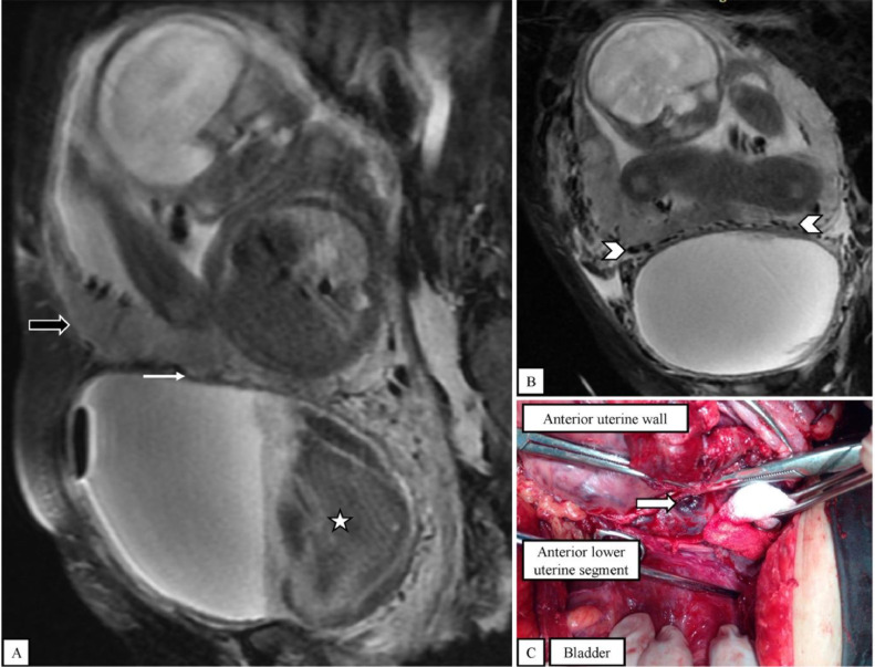
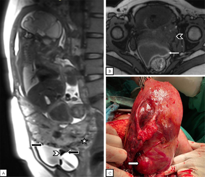
Placenta accreta spectrum (PAS) is defined as abnormal placental adherence or invasion of the myometrium or extrauterine organs. This case series will analyze MRI findings and PAS grading, in addition to emergency situations like massive hematuria and placental invasion with rupture. We report 5 cases of pregnant women with placenta previa with suspected PAS. MRI revealed 1 case of placenta accreta, one case of placenta increta, and 3 cases of placenta percreta. Two cases were emergency situations. All cases were managed with cesarean section. PAS is frequently related to severe obstetric hemorrhage associated with high maternal morbidity and mortality, making diagnosis and management challenging. Ultrasound is the initial diagnostic modality for PAS. Although ultrasound is preferred for PAS diagnosis, MRI provides an effective modality for the analysis of the depth of placental invasion and can be helpful in emergency situations.

Keywords: Diagnostic; Emergency; MRI; Placenta accreta spectrum.


Figures

Fig 1
Fig. 1
(A) Sagittal T2-weighted fat-saturated MR image shows an abnormal, heterogenous placenta with a rounded edge (star) and dark intraplacental bands (thin white arrows). There is mildly increased vascularity at the placental-myometrial interface (thick black arrow). (B) Cesarean section with hysterectomy shows minimal neovascularity and no placental tissue is seen invading the surface of the uterus (arrowhead). (C) Histopathological examination shows placental villi attached to the endometrium (thick white arrows).
Fig 2
Fig. 2
(A) Sagittal T2-weighted MR image shows an anteroinferior placental bulge into the myometrium (thick black arrow) and dark intraplacental bands (thin white arrow). (B) Cesarean section with hysterectomy shows a placental bulge with purple/blue coloring and multiple hypervascularity (arrowhead). No placental tissue is seen invading the surface of the uterus. (C) Histopathological examination shows placental villi invading the myometrium (thick white arrow).
Fig 3
Fig. 3
(A) Sagittal T2-weighted MR image shows the placental bulge into the uterine serosa, focal disruption of the myometrium, and loss of the retroplacental dark line (thick black arrow). There is also shortening of the cervix (thin white arrow). An abnormal, heterogenous placenta with lumpy contour, a rounded edge, and dark intraplacental bands are also seen (star). (B) Cesarean section with hysterectomy shows placental tissue invading the surface of the uterus (arrowhead). (C) Histopathological examination shows placental villi invading the uterine serosa (thick white arrows).
Fig 4
Fig. 4
(A) Sagittal T2-weighted fat-saturated MR image shows the placental bulge with loss of the retroplacental dark line (thick black arrow). There is a massive blood clot inside the bladder (star). The vesicouterine fat plane is still effective (thin white arrow). (B) Coronal T2-weighted fat-saturated MR image shows increased vascularity between the bladder and the uterus (arrowheads). (C) Cesarean section with hysterectomy shows placental tissue invading the surface of the uterus, without bladder infiltration (thick white arrow).
Fig 5
Fig. 5
(A) Sagittal T2-weighted MR image shows placental invasion to the bladder (thick black arrows). There is placental invasion of the S2 uterine sector (star) and a blood clot is seen inside the bladder (white arrowhead). (B) Axial T1-weighted MR image shows placental invasion with rupture (thin white arrow) and invasion to the lower-left parametrium (black arrowhead). (C) Cesarean section with hysterectomy shows placental tissue invading the surface of the bladder and left parametrium (thick white arrow).

Similar articles

Cited by

References

    1. Morlando M., Collins S. Placenta accreta spectrum disorders: challenges, risks, and management strategies. Int J Womens Health. 2020;12:1033–1045. doi: 10.2147/IJWH.S224191. Nov 10. - DOI - PMC - PubMed
    1. Wu S., Kocherginsky M., Hibbard J.U. Abnormal placentation: twenty-year analysis. Am J Obstet Gynecol. 2005;192(5):1458–1461. doi: 10.1016/j.ajog.2004.12.074. May. - DOI - PubMed
    1. Grechukhina O., Deshmukh U., Fan L., Kohari K., Abdel-Razeq S., Bahtiyar M.O., et al. Cesarean scar pregnancy, incidence, and recurrence: five-year experience at a single tertiary care referral center. Obstet Gynecol. 2018;132(5) doi: 10.1097/AOG.0000000000002940. - DOI - PubMed
    1. Aryananda R.A., Akbar A., Wardhana M.P., Gumilar K.E., Wicaksono B., Ernawati E., et al. New three-dimensional/four-dimensional volume rendering imaging software for detecting the abnormally invasive placenta. J Clin Ultrasound. 2019;47(1) doi: 10.1002/jcu.22641. - DOI - PubMed
    1. Nieto-Calvache A.J., Palacios-Jaraquemada J.M., Osanan G., Cortes-Charry R., Aryananda R.A., Bangal V.B., et al. Lack of experience is a main cause of maternal death in placenta accreta spectrum patients. Acta Obstet Gynecol Scand. 2021;100(8):1445–1453. doi: 10.1111/aogs.14163. Aug 1. - DOI - PubMed

Publication types

LinkOut - more resources