Case Reports
. 2022 Mar 28:77:103511.
doi: 10.1016/j.amsu.2022.103511. eCollection 2022 May.

Acute disseminated encephalitis (ADEM) as the first presentation of COVID-19; a case report

Affiliations
Case Reports

Acute disseminated encephalitis (ADEM) as the first presentation of COVID-19; a case report

Sara Esmaeili et al. Ann Med Surg (Lond). .

Abstract

Introduction: and importance: Neurological ailments are reported during and after SARS-COV-2 infection.

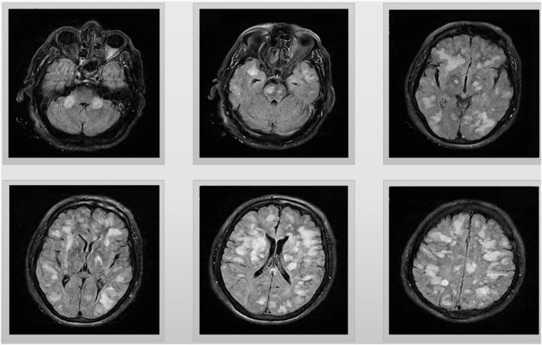
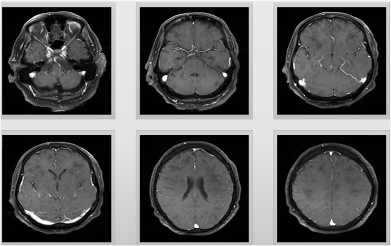
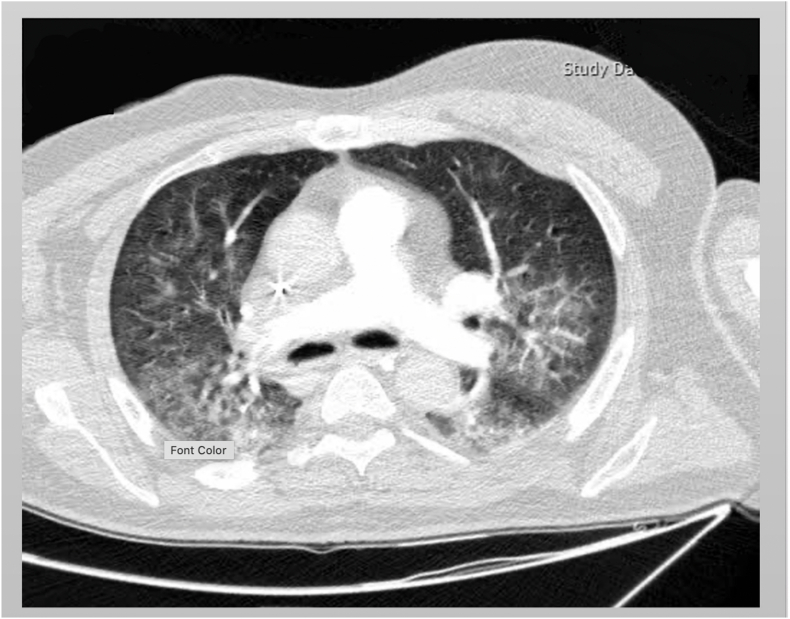
Case presentation: We report a 67-year-old Iranian man with COVID-19 infection and Acute Disseminated Encephalomyelitis (ADEM) whose neurological symptoms appeared before clinical and radiological pulmonary manifestations.

Clinical discussion: COVID-19 can cause neurological complication without entering the CNS via para infectious inflammatory mechanisms.

Conclusions: This report shows that ADEM might be among primary presentations of COVID-19.

Keywords: ADEM; Acute disseminated encephalomyelitis; Brain; COVID-19; Central nervous system; Neurologic.


Conflict of interest statement

The authors deny any conflict of interest in any terms or by any means during the study.

Figures

Fig. 1
Fig. 1
T2 flare views, axial magnetic resonance imaging (MRI), showing multifocal T2 hyperintense lesions with sparse enhancement on bilateral cerebral hemispheres, para ventricular and subcortical white matter, middle cerebellar peduncles, centrum semiovale, corpus callosum, thevbasal ganglia, thalami, midbrain and brain stem.
Fig. 2
Fig. 2
T1 postcontrast (gadolinium) views, axial magnetic resonance imaging (MRI), showing multifocal T2 hyperintense lesions with sparse enhancemen on bilateral cerebral hemispheres para ventricular and subcortical white matter, middle cerebellar peduncles, centrum semiovale, corpus callosum, thevbasal ganglia, thalami, midbrain and brain stem.
Fig. 3
Fig. 3
Chest CT scan shown ground glass opacities on the right middle lobe. There is also visible the reticular pattern on both lungs' fields.

Similar articles

Cited by

References

    1. Guan W-j, Ni Z-y, Hu Y., Liang W-h, Ou C-q, He J-x, et al. Clinical characteristics of coronavirus disease 2019. China. 2020;382(18):1708–1720. - PMC - PubMed
    1. Baig A.M., Khaleeq A., Ali U., Syeda H. Evidence of the COVID-19 virus targeting the CNS: tissue distribution, host–virus interaction, and proposed neurotropic mechanisms. ACS Chem. Neurosci. 2020;11(7):995–998. - PubMed
    1. Ellul M.A., Benjamin L., Singh B., Lant S., Michael B.D., Easton A., et al. Neurological associations of COVID-19. Lancet Neurol. 2020;19(9):767–783. - PMC - PubMed
    1. Aghagoli G., Gallo Marin B., Katchur N.J., Chaves-Sell F., Asaad W.F., Murphy S.A. Neurological involvement in COVID-19 and potential mechanisms: a review. Neurocritical Care. 2020:1–10. - PMC - PubMed
    1. Mao L., Jin H., Wang M., Hu Y., Chen S., He Q., et al. Neurologic manifestations of hospitalized patients with coronavirus disease 2019 in wuhan, China. JAMA Neurol. 2020;77(6):683–690. - PMC - PubMed

Publication types