

Incremental Detection of Severe Congenital Heart Disease by Fetal Echocardiography Following a Normal Second Trimester Ultrasound Scan in Québec, Canada
- PMID: 35369710
- PMCID: PMC9015032
- DOI: 10.1161/CIRCIMAGING.121.013796
Incremental Detection of Severe Congenital Heart Disease by Fetal Echocardiography Following a Normal Second Trimester Ultrasound Scan in Québec, Canada
Abstract
Background: The benefit of fetal echocardiograms (FE) to detect severe congenital heart diseases (SCHD) in the setting of a normal second-trimester ultrasound is unclear. We aimed to assess whether the increase in SCHD detection rates when FE are performed for risk factors in the setting of a normal ultrasound was clinically significant to justify the resources needed.
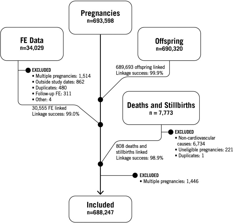
Methods: This is a multicenter, population-based, retrospective cohort study, including all singleton pregnancies and offspring in Quebec (Canada) between 2007 and 2015. Administrative health care data were linked with FE clinical data to gather information on prenatal diagnosis of CHD, indications for FE, outcomes of pregnancy and offspring, postnatal diagnosis of CHD, cardiac interventions, and causes of death. The difference between the sensitivity to detect SCHD with and without FE for risk factors was calculated using generalized estimating equations with a noninferiority margin of 5 percentage points.
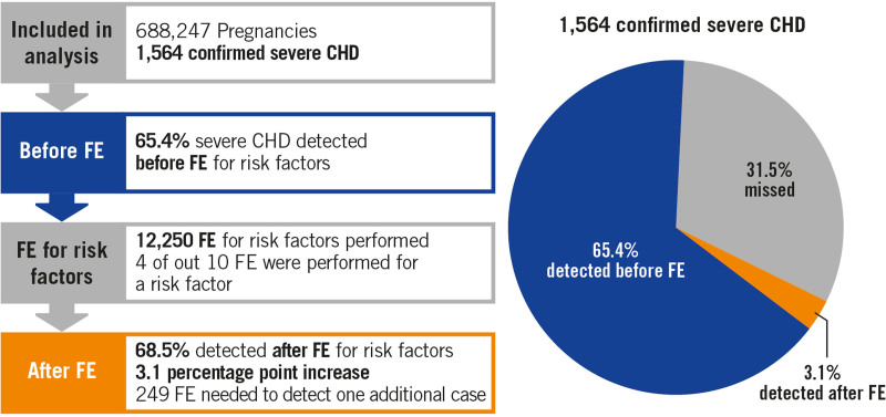
Results: A total of 688 247 singleton pregnancies were included, of which 30 263 had at least one FE. There were 1564 SCHD, including 1071 that were detected prenatally (68.5%). There were 12 210 FE performed for risk factors in the setting of a normal second-trimester ultrasound, which led to the detection of 49 additional cases of SCHD over 8 years. FE referrals for risk factors increased sensitivity by 3.1 percentage points (95% CI, 2.3-4.0; P<0.0001 for noninferiority).
Conclusions: In the setting of a normal second-trimester ultrasound, adding a FE for risk factors offered low incremental value to the detection rate of SCHD in singleton pregnancies. The current ratio of clinical gains versus the FE resources needed to screen for SCHD in singleton pregnancies with isolated risk factors does not seem favorable. Further studies should evaluate whether these resources could be better allocated to increase SCHD sensitivity at the ultrasound level, and to help decrease heterogeneity between regions, institutions and operators.
Keywords: congenital heart disease; echocardiography; pregnancy; prenatal diagnosis; risk factors.
Figures
Comment in
-
What Does Fetal Echocardiography Add Beyond the Anomaly Scan?Circ Cardiovasc Imaging. 2022 Apr;15(4):e014168. doi: 10.1161/CIRCIMAGING.122.014168. Epub 2022 Apr 4. Circ Cardiovasc Imaging. 2022. PMID: 35369699 No abstract available.
References
-
- Rocha LA, Araujo Júnior E, Rolo LC, Barros FS, da Silva KP, Leslie AT, Nardozza LM, Moron AF. Prenatal detection of congenital heart diseases: one-year survey performing a screening protocol in a single reference center in Brazil. Cardiol Res Pract. 2014;2014:175635. doi: 10.1155/2014/175635 - PMC - PubMed
-
- Rychik J, Ayres N, Cuneo B, Gotteiner N, Hornberger L, Spevak PJ, Van Der Veld M. American Society of Echocardiography guidelines and standards for performance of the fetal echocardiogram. J Am Soc Echocardiogr. 2004;17:803–810. doi: 10.1016/j.echo.2004.04.011 - PubMed
-
- Donofrio MT, Moon-Grady AJ, Hornberger LK, Copel JA, Sklansky MS, Abuhamad A, Cuneo BF, Huhta JC, Jonas RA, Krishnan A, et al. ; American Heart Association Adults With Congenital Heart Disease Joint Committee of the Council on Cardiovascular Disease in the Young and Council on Clinical Cardiology, Council on Cardiovascular Surgery and Anesthesia, and Council on Cardiovascular and Stroke Nursing. Diagnosis and treatment of fetal cardiac disease: a scientific statement from the American Heart Association. Circulation. 2014;129:2183–2242. doi: 10.1161/01.cir.0000437597.44550.5d - PubMed
-
- Finneran MM, Ware CA, Kiefer MK, Buschur EO, Foy PM, Thung SF, Landon MB, Gabbe SG. The accuracy and cost-effectiveness of selective fetal echocardiography for the diagnosis of congenital heart disease in patients with pregestational diabetes stratified by hemoglobin A1c. Am J Perinatol. 2019;36:1216–1222. doi: 10.1055/s-0039-1685490 - PubMed
-
- Garg S, Sharma P, Sharma D, Behera V, Durairaj M, Dhall A. Use of fetal echocardiography for characterization of fetal cardiac structure in women with normal pregnancies and gestational diabetes mellitus. J Ultrasound Med. 2014;33:1365–1369. doi: 10.7863/ultra.33.8.1365 - PubMed
Publication types
MeSH terms
Grants and funding
LinkOut - more resources
Full Text Sources
Other Literature Sources
Medical
