[Clinical and radiological findings for the new multisystem inflammatory syndrome in children associated with COVID-19]
- PMID: 35370316
- PMCID: PMC7951883
- DOI: 10.1016/j.rx.2021.03.001
[Clinical and radiological findings for the new multisystem inflammatory syndrome in children associated with COVID-19]
Abstract
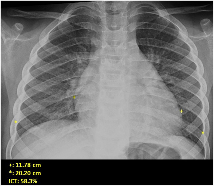
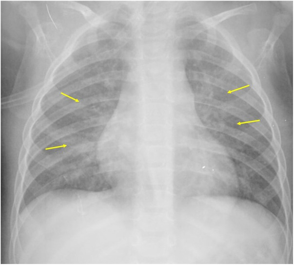
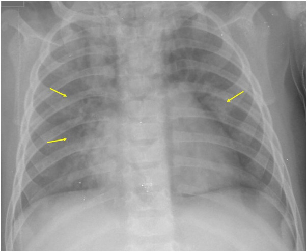
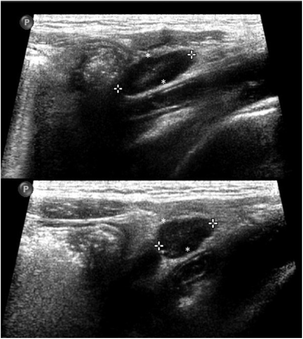
The World Health Organization defines the multisystem inflammatory syndrome in children (MIS-C) as a new syndrome reported in patients aged < 19 years old who have a history of exposure to SARS-CoV-2. The onset of this syndrome is characterized by persistent fever that is associated with lethargy, abdominal pain, vomiting and/or diarrhea, and, less frequently, rash and conjunctivitis. The course and severity of the signs and symptoms vary; in some children, MIS-C worsens rapidly and can lead to hypotension, cariogenic shock, or even damage to multiple organs. The characteristic laboratory findings are elevated markers of inflammation and heart dysfunction. The most common radiological findings are cardiomegaly, pleural effusion, signs of heart failure, ascites, and inflammatory changes in the right iliac fossa. In the context of the current COVID-19 pandemic, radiologists need to know the clinical, laboratory, and radiological characteristics of this syndrome to ensure the correct diagnosis.
El síndrome inflamatorio multisistémico pediátrico vinculado a la COVID-19 (SIM-PedS) es, según la Organización Mundial de la Salud, un nuevo síndrome descrito en pacientes menores de 19 años con historia previa de exposición a SARS-CoV-2. La presentación inicial de este síndrome se caracteriza por fiebre persistente que asocia debilidad, dolor abdominal, vómitos y/o diarrea. Menos frecuentemente los pacientes pueden presentar también erupción cutánea y conjuntivitis. El cuadro clínico tiene expresividad y evolución variables, por lo que algunos pacientes pediátricos afectados pueden empeorar rápidamente, desarrollando desde hipotensión y shock cardiogénico a daño multiorgánico. Los hallazgos analíticos característicos del síndrome consisten en elevación de marcadores inflamatorios y disfunción cardíaca. Los hallazgos radiológicos más frecuentes son cardiomegalia, derrame pleural, signos de insuficiencia cardíaca, ascitis y cambios inflamatorios en la fosa ilíaca derecha. En la pandemia actual por COVID-19 es necesario que el radiólogo conozca las características clínico-analíticas y radiológicas de este síndrome para realizar un correcto diagnóstico.
Keywords: COVID-19; Multisystem inflammatory syndrome in children; SARS-CoV-2.
© 2021 SERAM. Published by Elsevier España, S.L.U. All rights reserved.
Figures


Similar articles
-
Clinical and radiological findings for the new multisystem inflammatory syndrome in children associated with COVID-19.Radiologia (Engl Ed). 2021 Jul-Aug;63(4):334-344. doi: 10.1016/j.rxeng.2021.03.005. Epub 2021 Jun 5. Radiologia (Engl Ed). 2021. PMID: 34246424 Free PMC article.
-
Hematuria as an Early Sign of Multisystem Inflammatory Syndrome in Children: A Case Report of a Boy With Multiple Comorbidities and Review of Literature.Front Pediatr. 2021 Oct 27;9:760070. doi: 10.3389/fped.2021.760070. eCollection 2021. Front Pediatr. 2021. PMID: 34778150 Free PMC article.
-
Imaging Findings in Multisystem Inflammatory Syndrome in Children (MIS-C) Associated With Coronavirus Disease (COVID-19).AJR Am J Roentgenol. 2021 Feb;216(2):507-517. doi: 10.2214/AJR.20.24032. Epub 2020 Jul 29. AJR Am J Roentgenol. 2021. PMID: 32755212
-
COVID-19 Associated Multisystem Inflammatory Syndrome: A Systematic Review and Meta-analysis.Iran J Allergy Asthma Immunol. 2020 Dec 19;19(6):570-588. doi: 10.18502/ijaai.v19i6.4927. Iran J Allergy Asthma Immunol. 2020. PMID: 33463127
-
Similarities and differences between multiple inflammatory syndrome in children associated with COVID-19 and Kawasaki disease: clinical presentations, diagnosis, and treatment.World J Pediatr. 2021 Aug;17(4):335-340. doi: 10.1007/s12519-021-00435-y. Epub 2021 May 20. World J Pediatr. 2021. PMID: 34013488 Free PMC article. Review.
References
Publication types
LinkOut - more resources
Full Text Sources
Miscellaneous